

Clear, labeled diagrams for the concepts students and researchers look up most — read one, or generate your own version with AI.
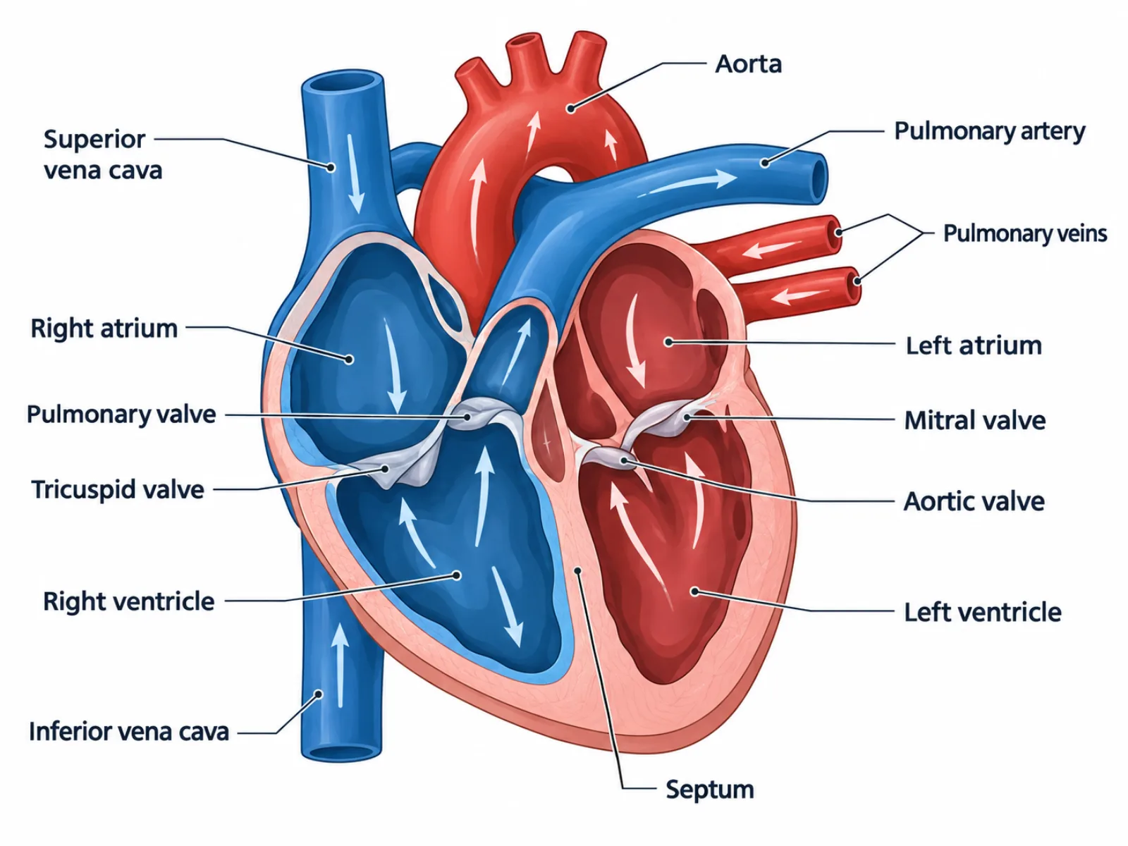
See how a heart diagram maps chambers, valves, and vessels to the path blood actually takes, and why the left/right layout trips up so many first-time readers.
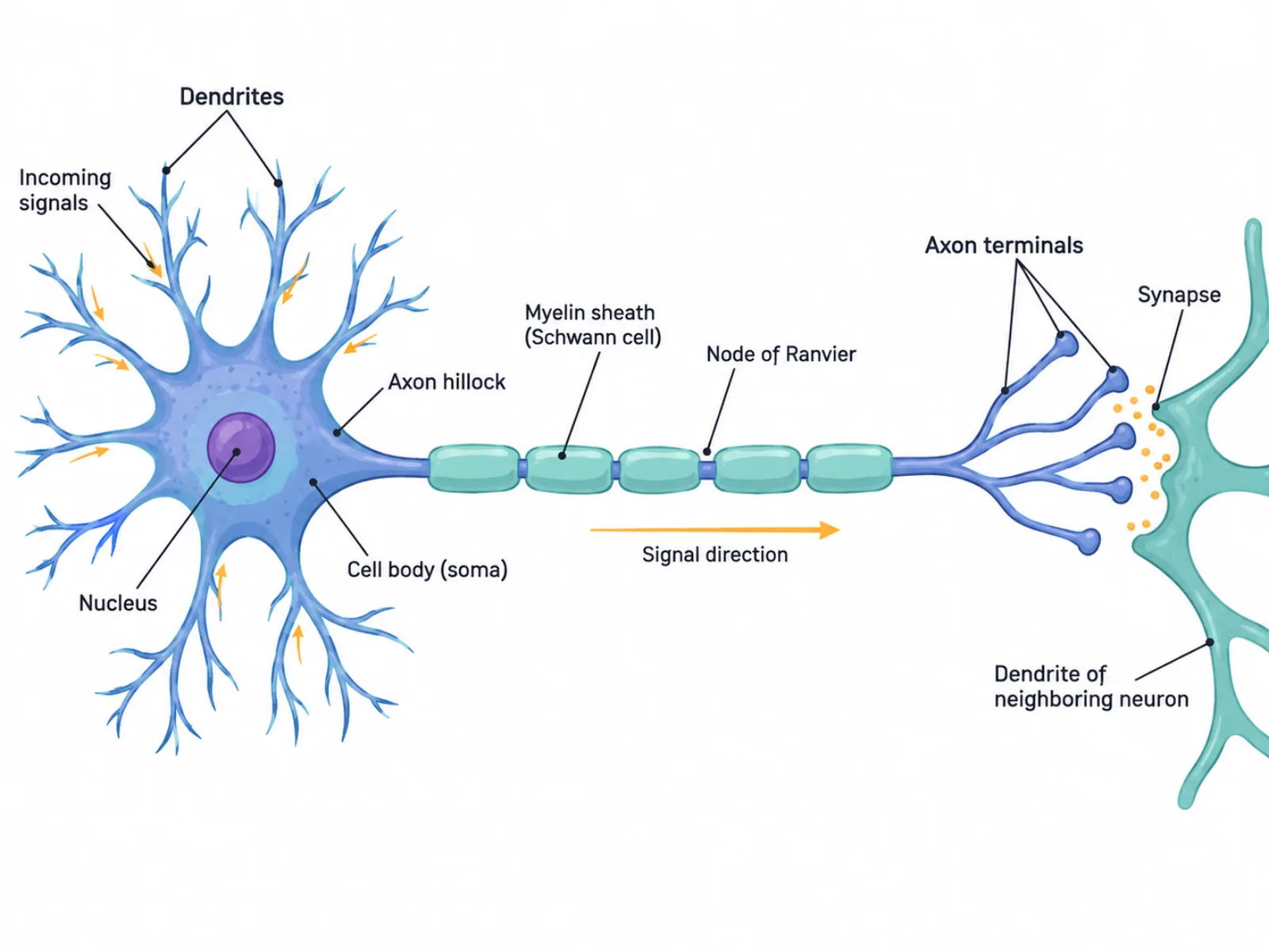
Break down a neuron diagram structure by structure, from dendrites to synaptic terminals, and learn why signal direction is the detail most diagrams get wrong.
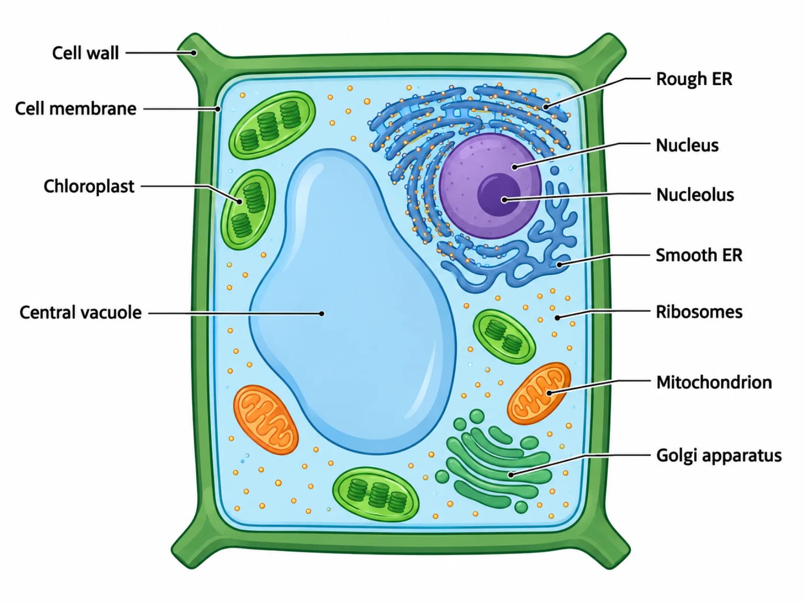
Understand every organelle in a plant cell diagram, why the cell wall forces its boxy shape, and how it differs structurally from an animal cell.
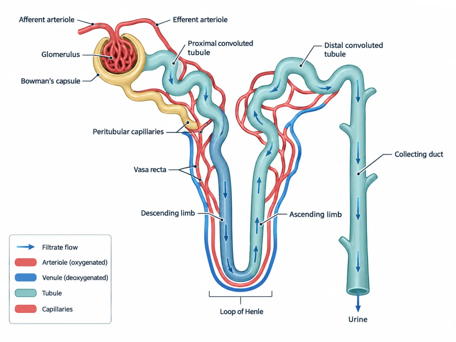
Trace filtrate through a nephron diagram from glomerulus to collecting duct, and learn why filtration, reabsorption, and secretion are three distinct steps.
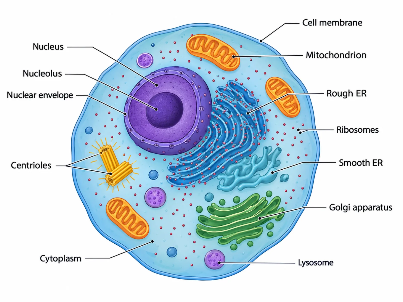
Identify every organelle in an animal cell diagram, understand why its outline is round rather than boxy, and see how it compares to a plant cell.
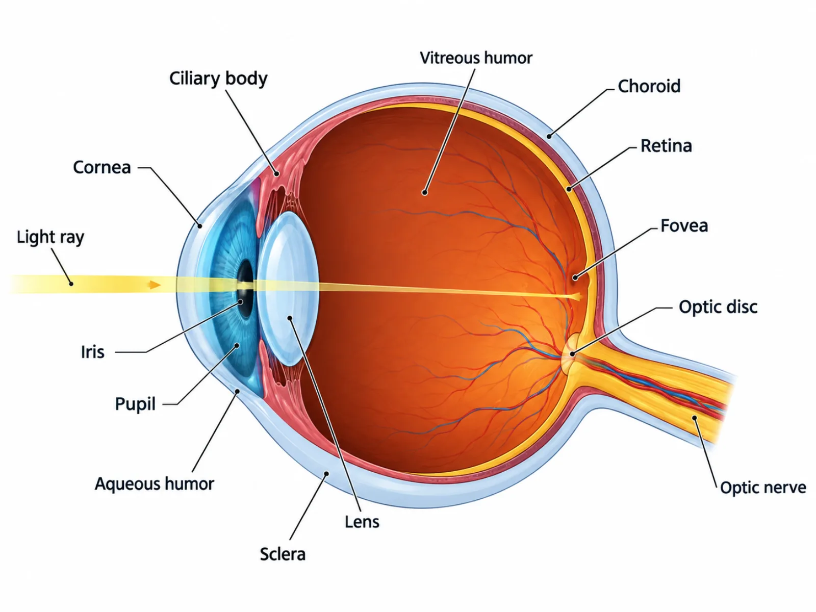
A labeled human eye diagram traces how light travels from cornea to retina, why the image lands upside down, and where the blind spot actually sits.
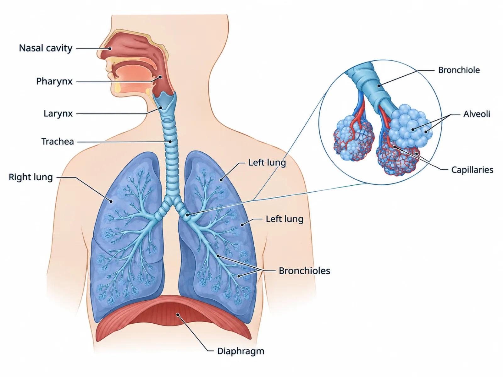
A respiratory system diagram guide to the airway from nose to alveoli, plus the common mix-up between organ-level breathing and cellular respiration.
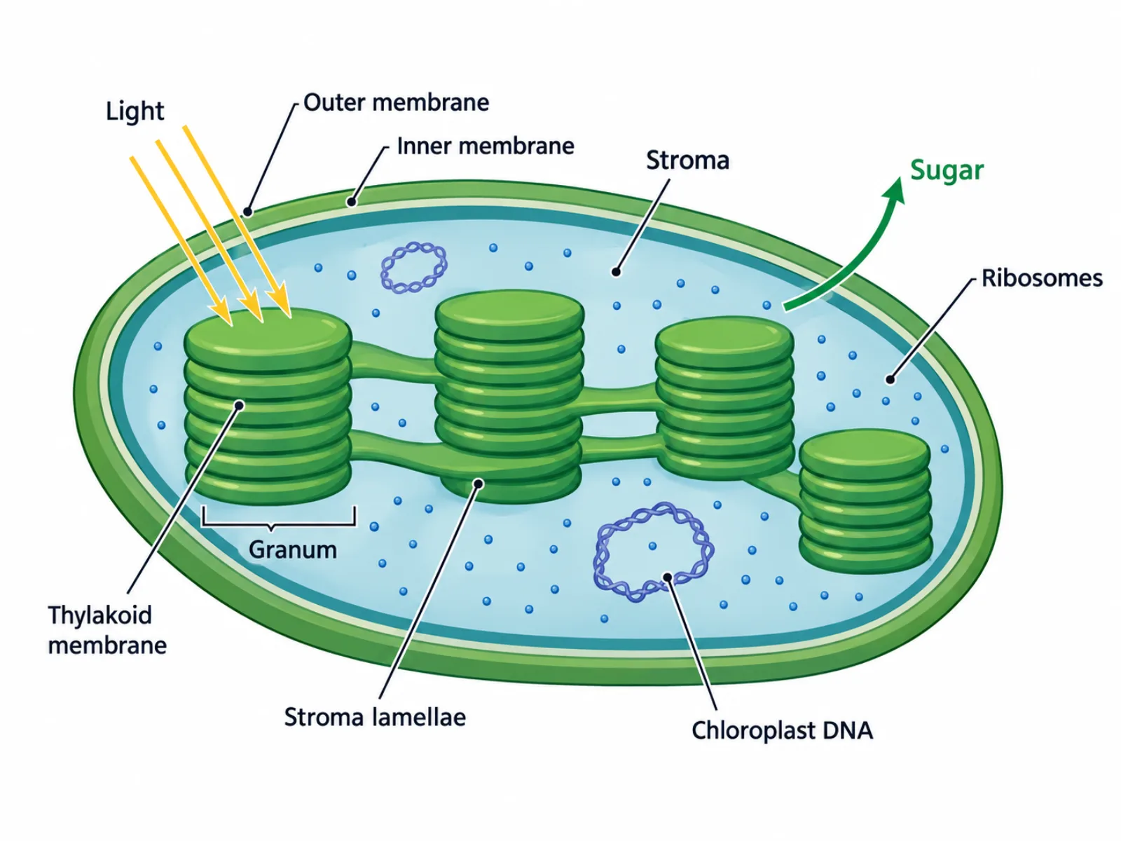
A chloroplast diagram maps the thylakoid stacks, stroma, and double membrane where photosynthesis happens, plus why chloroplasts share so much with mitochondria.
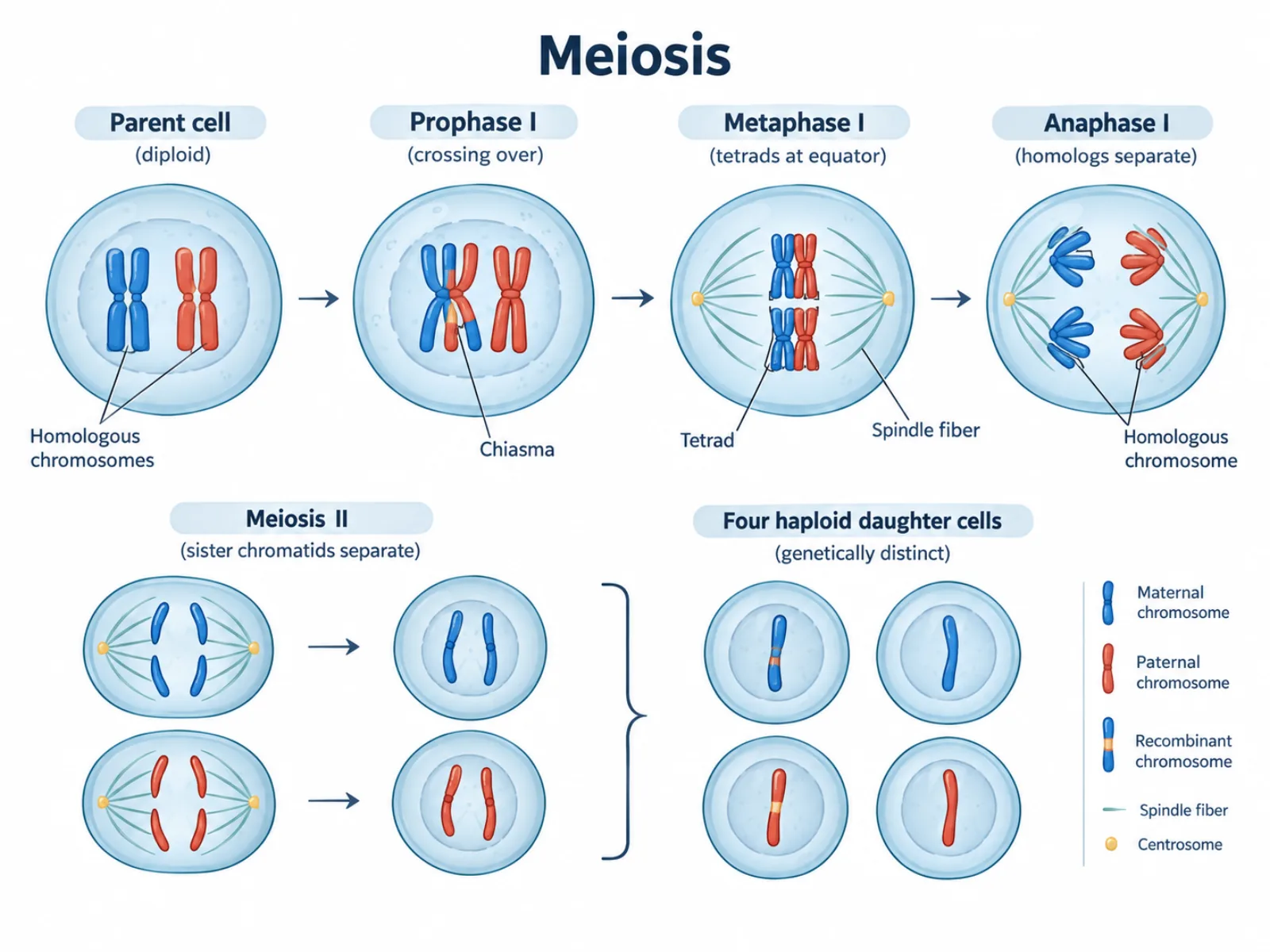
A meiosis diagram tracks how one cell becomes four unique gametes, marking the exact stage where homologous chromosomes and sister chromatids each separate.
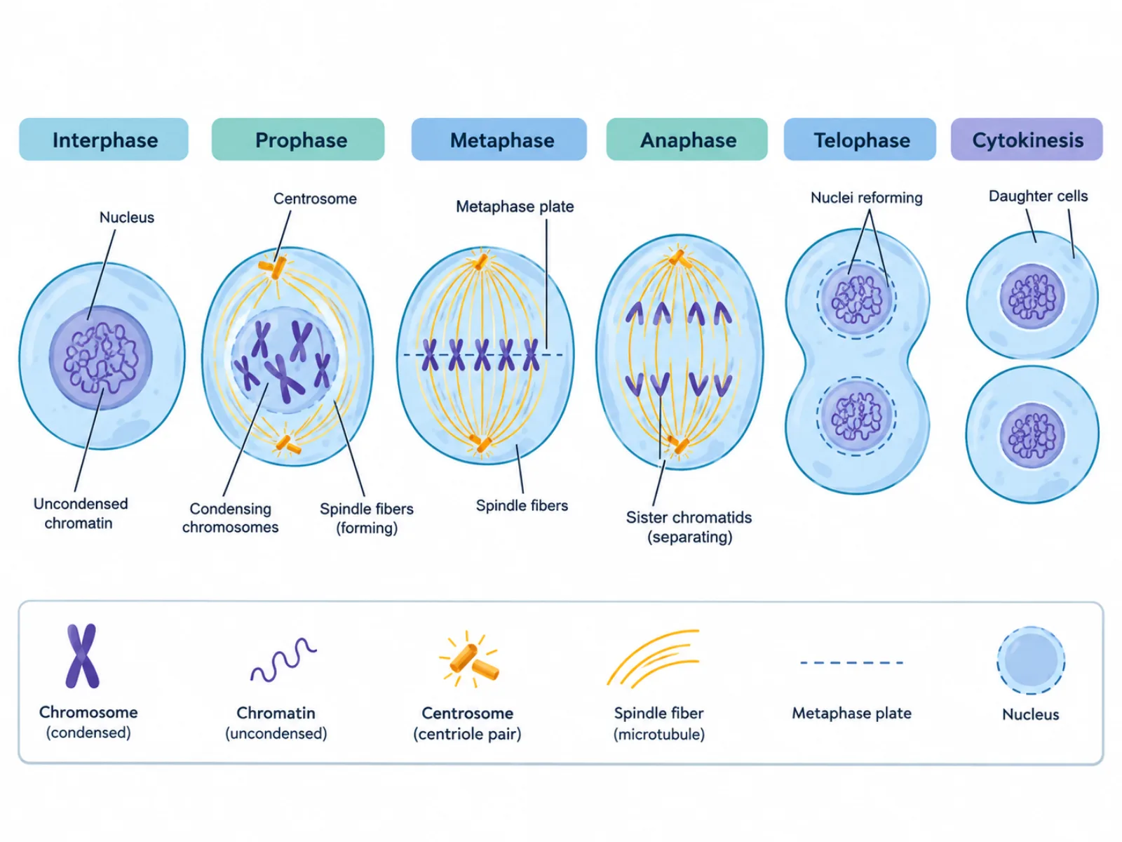
This mitosis diagram guide walks through each phase in order, clears up the chromosome-versus-chromatid mix-up, and contrasts the process with meiosis.
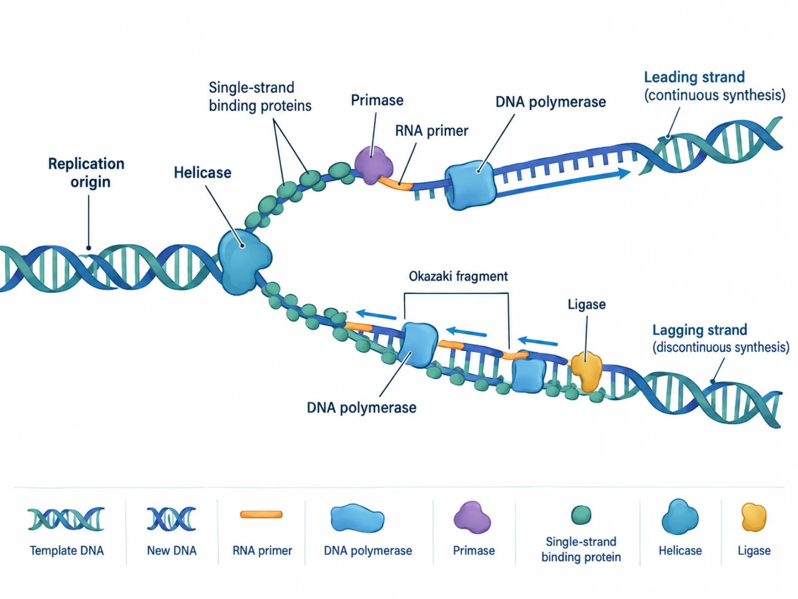
Trace how a replication fork opens, why one new strand runs continuously and the other in fragments, and which enzyme does which job — laid out step by step.
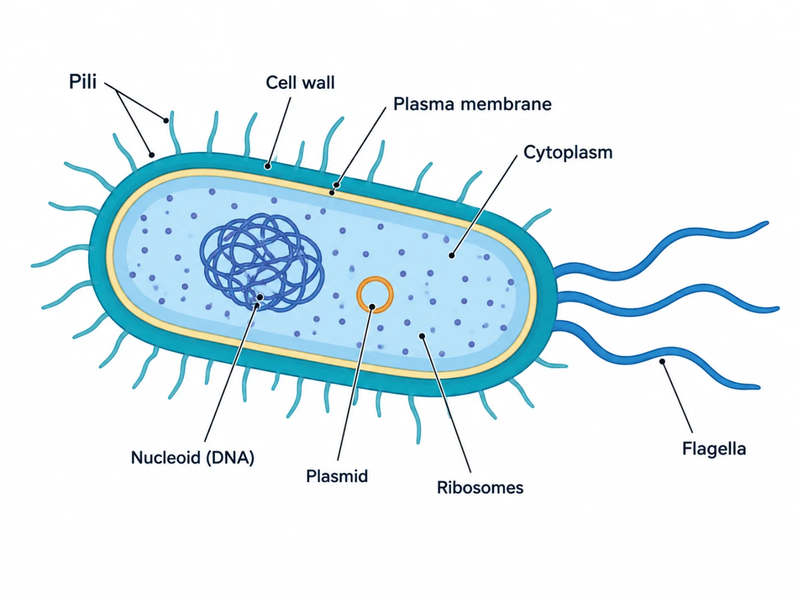
See why a prokaryotic cell diagram has no nucleus, what the nucleoid and plasmid actually are, and how each surface structure earns its place in the drawing.
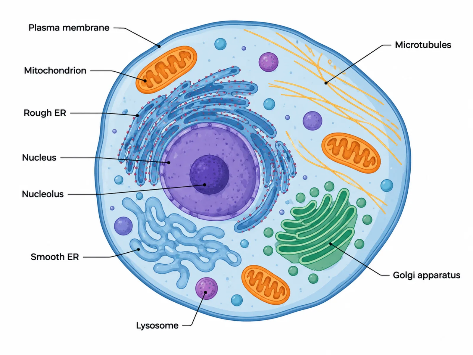
Understand what makes a eukaryotic cell diagram distinct — a true nucleus, an interconnected endomembrane system, and organelles no bacterial cell possesses.
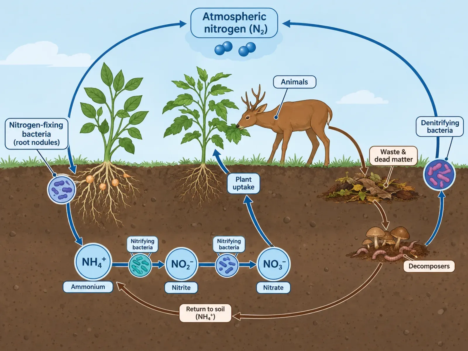
Follow nitrogen from air to soil to living things and back — the five processes a nitrogen cycle diagram must show and where readers usually go wrong.
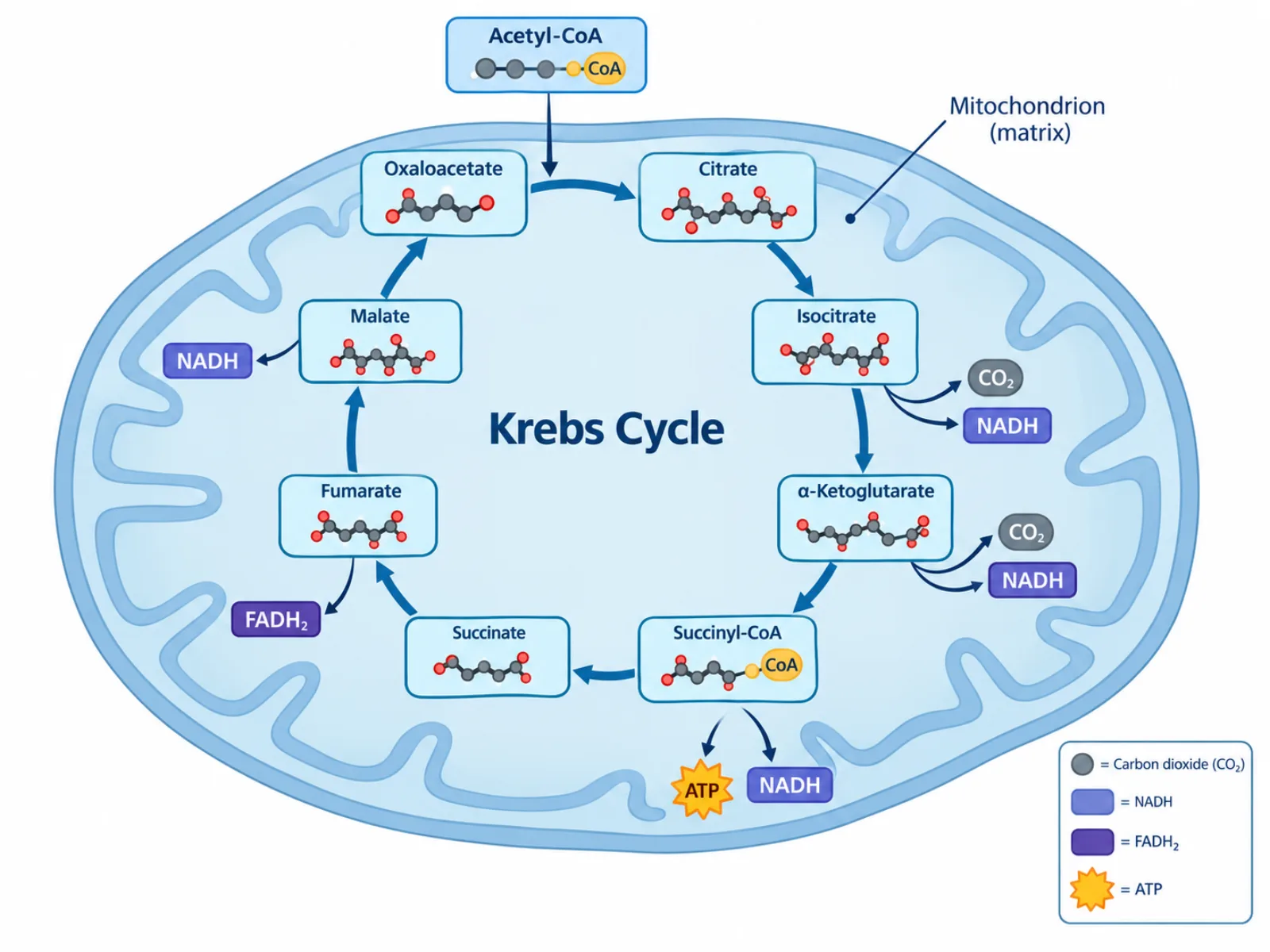
See where citrate forms, why oxaloacetate keeps reappearing, and what the Krebs cycle actually produces per turn — a step-by-step reading of the diagram.
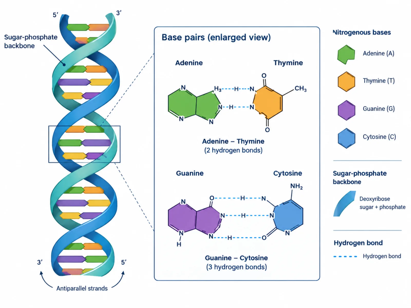
A visual breakdown of the antiparallel backbones, base-pairing rules, and grooves in a DNA structure diagram, plus the pairing mistakes most learners make.
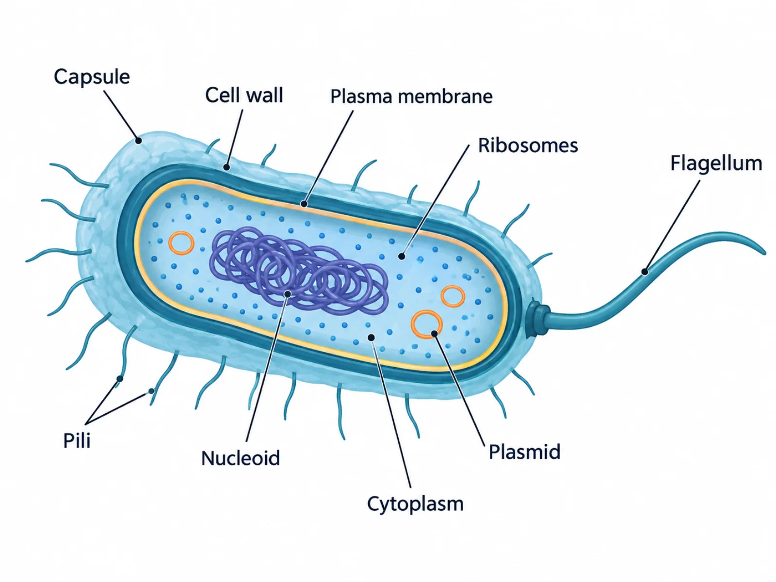
A guide to the structures in a labeled bacterial cell diagram, from the capsule and cell wall to plasmids and pili, plus the mix-ups that trip up learners.
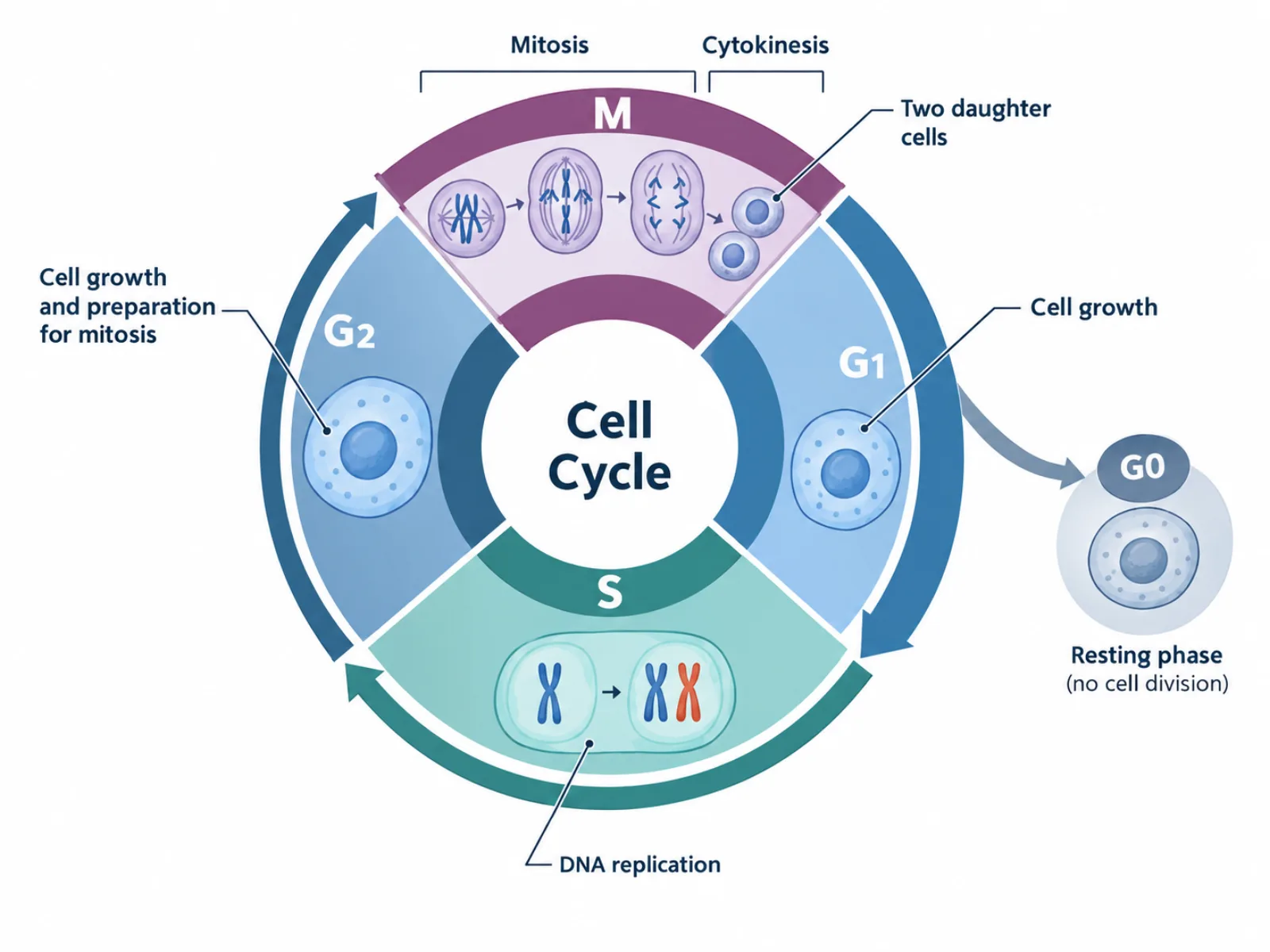
Why a cell cycle diagram is drawn as a circle, what each phase and checkpoint actually verifies, and the timing mistake that makes most first drafts inaccurate.
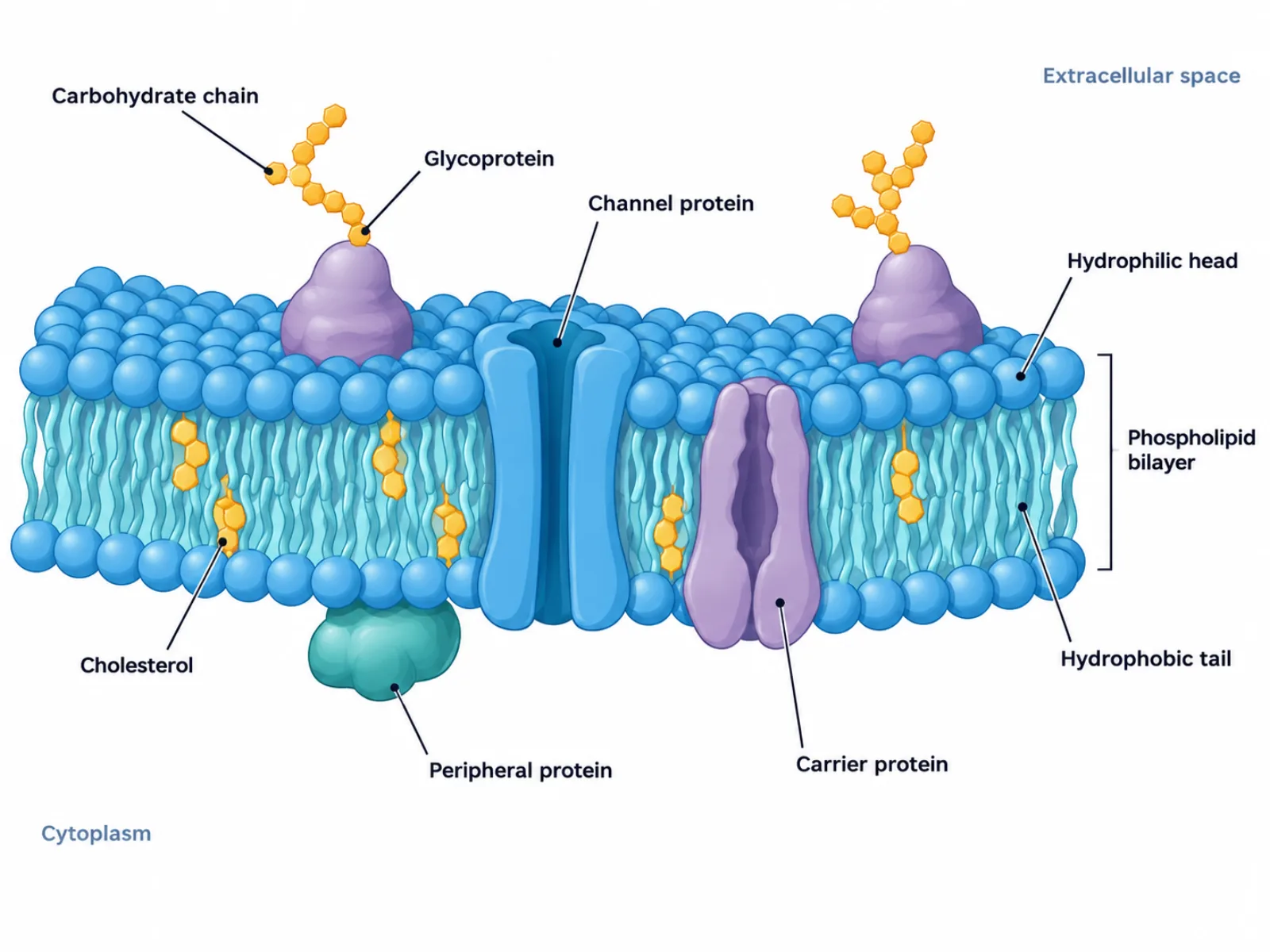
A labeled breakdown of the phospholipid bilayer, membrane proteins, and cholesterol in a cell membrane diagram, and why it isn't a rigid wall.
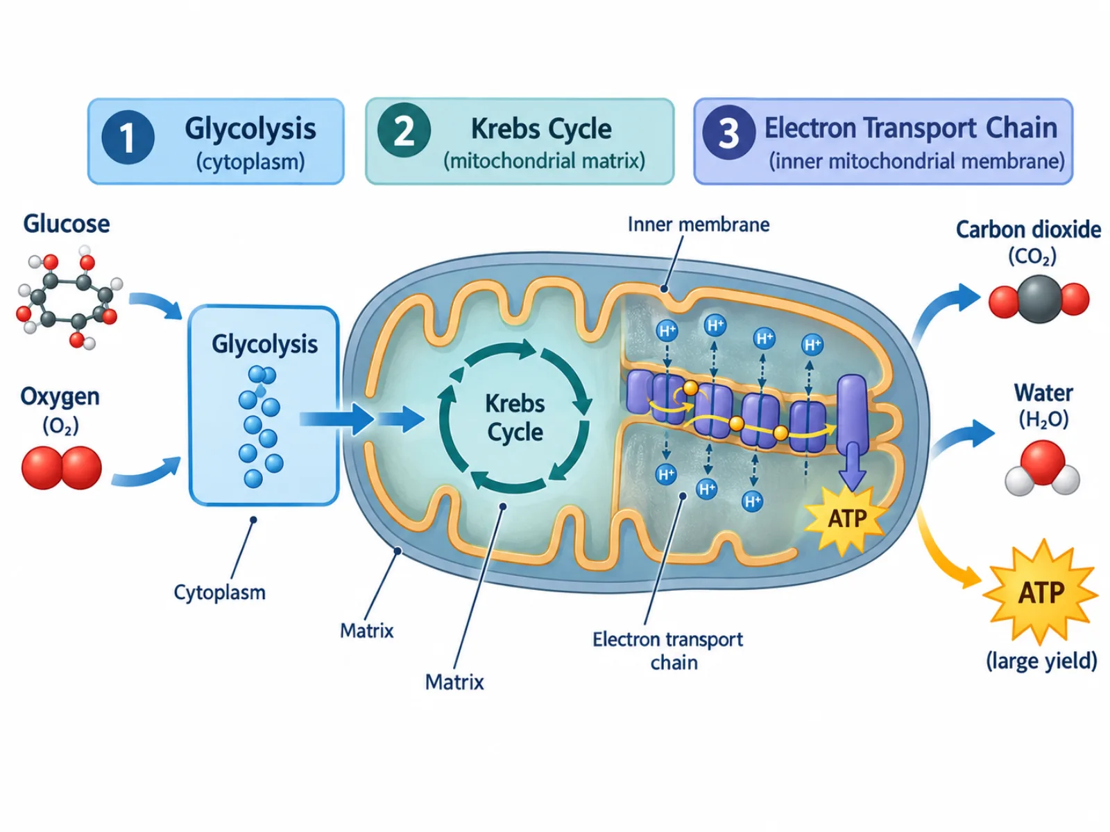
How a cellular respiration diagram traces glucose through three stages to ATP, why most ATP comes from the last stage, and how it differs from photosynthesis.