
Cell Membrane Diagram: The Fluid Mosaic Model Explained
A cell membrane diagram is a molecular close-up of the thin, selectively permeable boundary that surrounds every cell, illustrated according to the fluid mosaic model. Rather than showing the membrane as a simple outline, a proper cell membrane diagram zooms far enough in to depict individual phospholipids, embedded proteins, and cholesterol molecules, revealing the membrane as a dynamic, moving structure rather than a static wall. Because so much of a cell's behavior — what enters, what exits, what it recognizes as self or foreign — depends on this layer, a clearly labeled cell membrane diagram is essential for understanding nearly every other cellular process.
What the Cell Membrane Diagram Shows
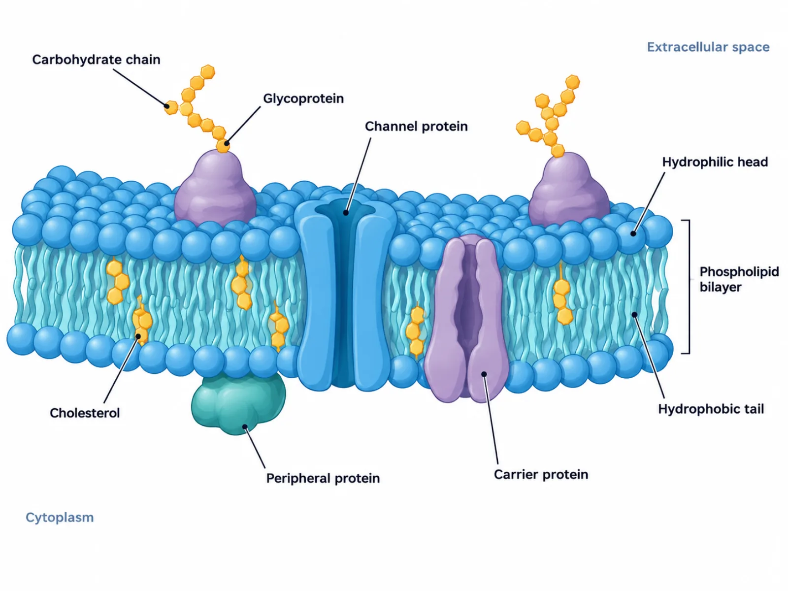
The diagram represents the fluid mosaic model: a double layer of phospholipids studded with a shifting arrangement of proteins, resembling a mosaic of different pieces scattered across a fluid background. The bilayer forms the structural foundation, oriented so that its water-attracting portions face outward on both sides and its water-repelling portions face each other in the middle, sandwiched between the two watery environments inside and outside the cell. Embedded within and attached to this bilayer are various proteins and cholesterol molecules, each contributing a different function — some regulate what crosses the membrane, others let the cell recognize its surroundings, and others simply hold the structure at the right level of flexibility.
The Labeled Parts of a Cell Membrane Diagram
- Phospholipid bilayer — two layers of phospholipids forming the membrane's basic structure, with hydrophilic heads facing outward and hydrophobic tails facing inward.
- Hydrophilic phosphate head — the water-attracting portion of each phospholipid, always facing the watery environment on either side of the membrane.
- Hydrophobic fatty acid tail — the water-repelling portion of each phospholipid, always facing inward, away from water.
- Integral membrane proteins — proteins that span the entire bilayer, including channel proteins and carrier proteins.
- Channel proteins — integral proteins that form pores allowing specific ions or molecules to pass passively through the membrane.
- Carrier proteins — integral proteins that change shape to actively transport specific molecules across the membrane.
- Peripheral proteins — proteins attached to only one surface of the membrane without spanning it, often involved in cell signaling or maintaining cell shape.
- Cholesterol — molecules wedged between the phospholipids that regulate membrane fluidity across a range of temperatures.
- Glycoproteins and glycolipids — proteins and lipids with attached carbohydrate chains, found only on the outer surface, functioning as identity markers for cell recognition.
How to Interpret and Draw the Diagram
A cell membrane diagram is best read as a cross-section, viewed from the side so both layers of the bilayer are visible stacked on top of each other, with the extracellular space above and the cytoplasm below. Every phospholipid should show its head pointing toward one of the two watery environments and its tail pointing toward the opposite phospholipid layer, never the reverse. Proteins should be distinguished by how far they extend: integral proteins fully cross both layers of the bilayer, while peripheral proteins touch only one surface. Carbohydrate chains, when present, should only ever extend from the outer-facing surface into the extracellular space, since their role in cell recognition depends on being visible to the outside environment. The overall layout should suggest movement and irregularity rather than a neat, repeating grid, since the model's defining feature is that its components drift and rearrange rather than sit in fixed positions.
Common Mistakes When Reading or Drawing a Cell Membrane Diagram
One of the most frequent errors is drawing carbohydrate chains on the inner, cytoplasmic surface of the membrane. Glycoproteins and glycolipids only ever appear on the outer surface, facing the extracellular space, since their function as recognition markers depends on being accessible to other cells and molecules outside. A second common mistake is treating the membrane as a rigid, static wall with fixed, unmoving components, when the fluid mosaic model specifically describes a structure whose phospholipids and proteins can drift laterally within the layer. A third mistake is confusing channel proteins with carrier proteins: channel proteins are passive pores that simply allow molecules through, while carrier proteins physically change shape to move molecules across, sometimes expending energy to do so — conflating the two misrepresents how selective transport across the membrane actually works.
Cell Membrane Diagram vs. Whole-Cell Diagram
A cell membrane diagram is often mistaken for part of a broader whole-cell diagram, but the two operate at very different scales of detail. A cell membrane diagram is a molecular close-up, detailed enough to show individual phospholipids, embedded proteins, and cholesterol arranged according to the fluid mosaic model. A whole animal- or plant-cell diagram, by contrast, shows that same membrane only as a single thin line encircling the entire cell, since its purpose is to depict the cell's overall shape and the arrangement of its internal organelles rather than the membrane's own molecular composition. Understanding which diagram is being asked for matters: a whole-cell diagram calls for a simple boundary line, while a cell membrane diagram calls for the full layered, labeled structure described above.