
Plant Cell Diagram: Labeled Organelles Guide
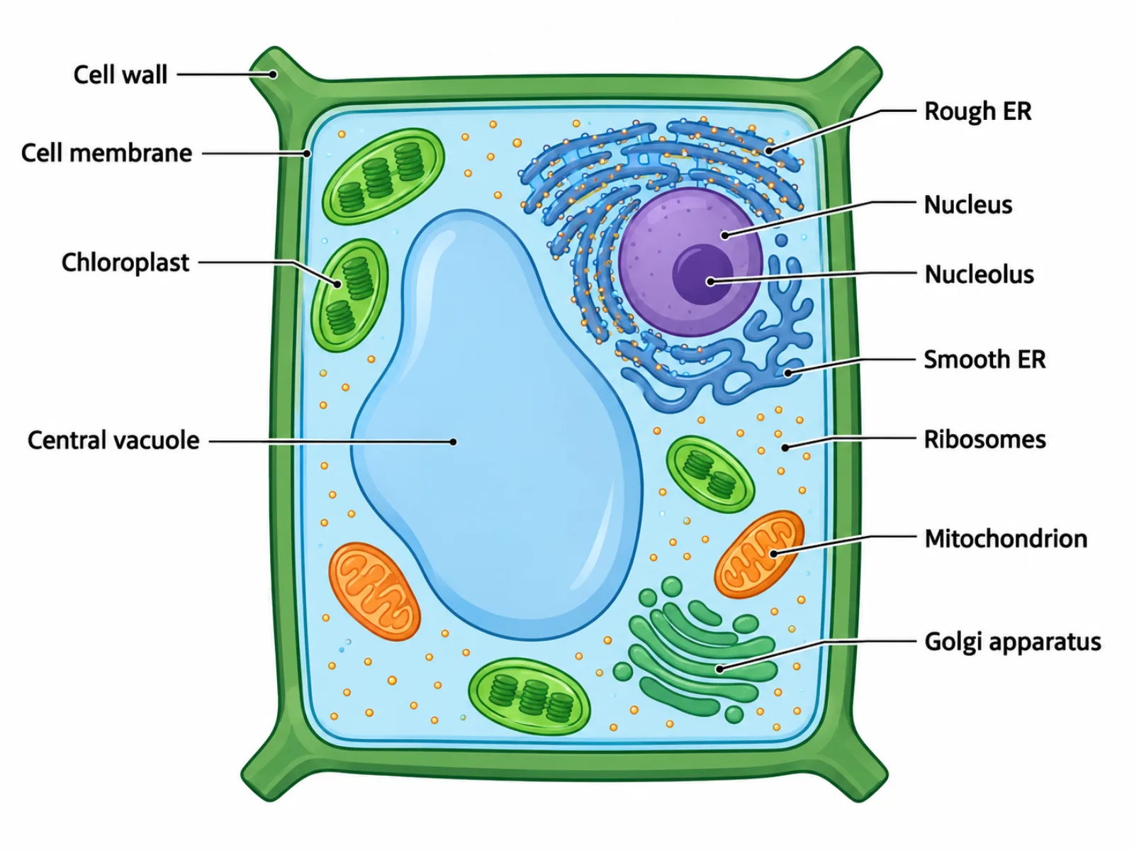
A plant cell diagram is a labeled cross-section of a typical plant cell showing the organelles and structures that let it carry out photosynthesis, maintain rigidity, and store nutrients. Unlike an animal cell diagram, a plant cell diagram has a distinctly angular, boxy outline because of the rigid wall surrounding the cell, and it dedicates a large portion of its interior to a single fluid-filled sac. Learning to read a plant cell diagram means recognizing not just what each organelle is called, but why its size and position reflect the specific demands of being a stationary, photosynthetic, and structurally supported cell.
What a Plant Cell Diagram Shows
The diagram represents the internal architecture of a single plant cell, built to capture energy from sunlight, hold its shape without a skeleton, and store water and nutrients efficiently. Every structure inside a plant cell diagram serves one of three broad purposes: generating or storing energy, maintaining the cell's shape and internal pressure, or manufacturing and processing proteins and other molecules. Because plant cells cannot move to find better conditions, many of their structures — the wall, the vacuole, the chloroplasts — are built for stability and self-sufficiency rather than mobility.
The Labeled Parts of a Plant Cell Diagram
- Cell wall — a rigid layer of cellulose outside the cell membrane that gives the cell its fixed shape and structural support.
- Cell membrane — the flexible barrier just inside the cell wall that controls what substances enter and exit the cell.
- Large central vacuole — stores water and nutrients and maintains turgor pressure that pushes against the cell wall, keeping the cell firm.
- Chloroplast — the site of photosynthesis, where light energy is converted into chemical energy stored in sugars.
- Nucleus with nucleolus — controls gene expression and directs the cell's overall activity; the nucleolus within it produces ribosomal components.
- Mitochondria — produce ATP through cellular respiration, supplying usable energy for the cell's activities.
- Rough endoplasmic reticulum — studded with ribosomes, it synthesizes proteins destined for secretion or membranes.
- Smooth endoplasmic reticulum — synthesizes lipids and helps process other molecules, lacking the ribosomes of its rough counterpart.
- Golgi apparatus — modifies, sorts, and packages proteins for delivery to their final destinations.
- Ribosomes — carry out protein synthesis, either freely in the cytoplasm or attached to the rough endoplasmic reticulum.
How to Interpret and Draw the Diagram
The most useful way to read a plant cell diagram is to notice how much of the interior a single structure occupies, because size in this diagram is not arbitrary — it reflects real proportions. The central vacuole should dominate the visual space, often taking up the majority of the cell's interior, with the nucleus and other organelles compressed toward the periphery as a result. The cell wall should be drawn as a distinct double line or thick border outside the cell membrane, making clear that it is a separate structure rather than just a thicker version of the membrane. Chloroplasts are usually scattered near the cell's edge, closer to the membrane, since they need access to light and gas exchange. When drawing the diagram, start with the outer wall to fix the cell's angular shape, then add the vacuole as the dominant internal feature before filling in the smaller organelles around it — working from largest, most defining structures to smallest mirrors how the cell's own proportions actually work.
Common Mistakes When Reading or Drawing a Plant Cell Diagram
A frequent mistake is drawing the cell with a rounded or irregular outline, as if it were an animal cell. Because the cellulose cell wall is rigid, real plant cells maintain a fixed, often rectangular or polygonal shape, and a diagram that rounds off the corners misrepresents this basic structural fact. Another common error is omitting the cell wall entirely or drawing the central vacuole too small relative to the rest of the cell; in a mature plant cell the vacuole can take up around 90 percent of total volume, so a vacuole sketched as a minor internal blob understates its actual dominance. A third mistake is assuming every plant cell diagram must include visible chloroplasts. Chloroplasts are only present in photosynthetic tissue, such as leaf cells, and are typically absent from root cells and other non-photosynthetic plant tissue, so a generic root cell diagram that includes chloroplasts is factually inaccurate.
Plant Cell Diagram vs. Animal Cell Diagram
The clearest way to understand a plant cell diagram is by direct contrast with an animal cell diagram. Plant cells have a rigid cell wall outside the membrane; animal cells have no wall at all, only the flexible membrane, which is why animal cells are drawn with a rounded or irregular outline instead of a fixed angular shape. Plant cells feature one large central vacuole that can dominate the cell's volume; animal cells instead contain only small vesicles scattered through the cytoplasm, none large enough to be called a central vacuole. Plant cells contain chloroplasts for photosynthesis in tissues exposed to light; animal cells never contain chloroplasts, since animals obtain energy by consuming other organisms rather than capturing sunlight directly. These three differences — wall, vacuole size, and chloroplasts — are the fastest way to tell the two diagram types apart at a glance, and they are not stylistic choices but direct consequences of how plant and animal cells actually function.