

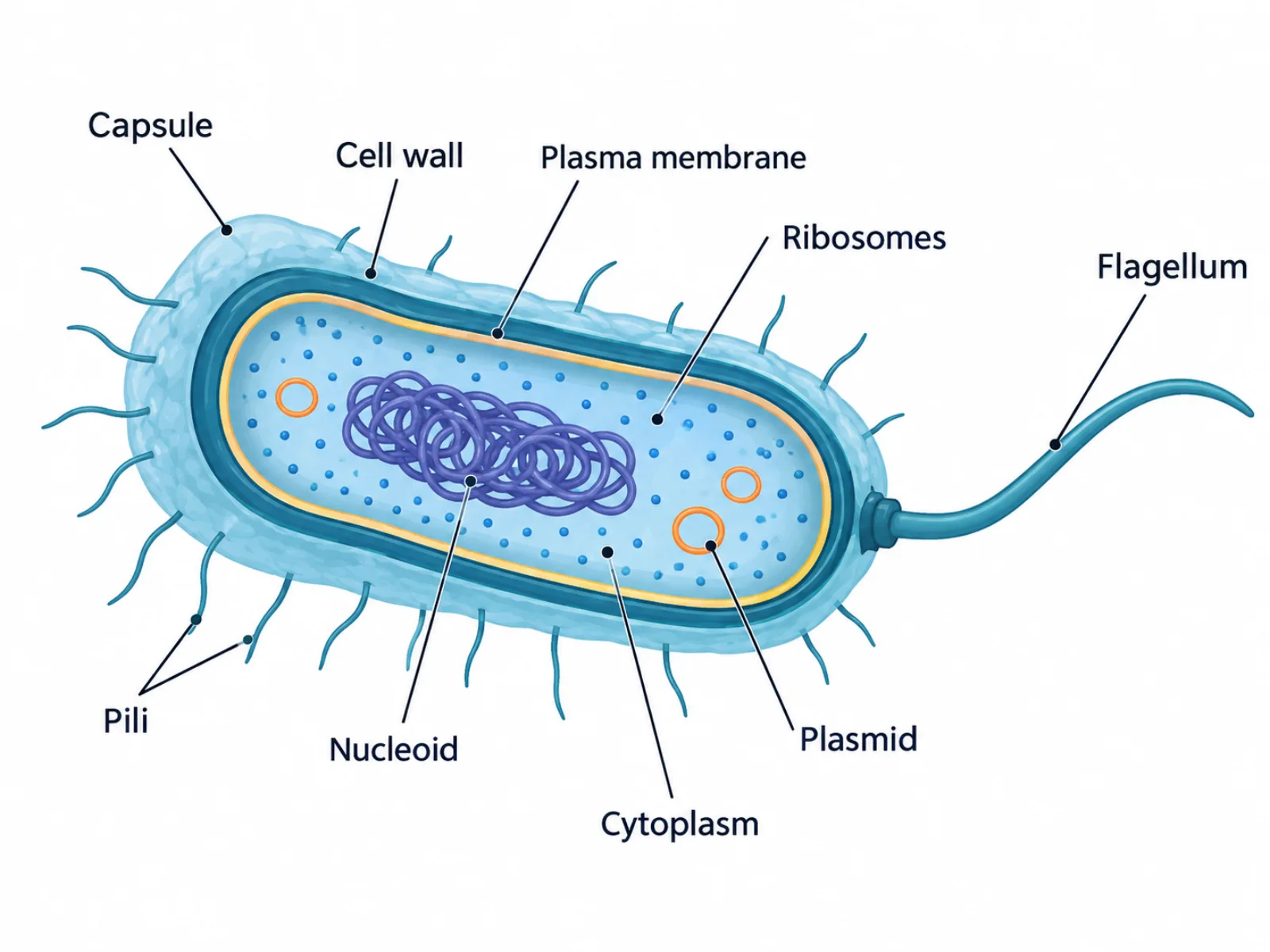
Bacterial Cell Diagram: Labeled Structures
A bacterial cell diagram is a labeled cross-section of a single bacterium, showing the structures that let this simple, single-celled organism survive, move, defend itself, and reproduce. Because bacteria lack the internal membrane-bound organelles found in animal or plant cells, a bacterial cell diagram looks comparatively sparse — but every structure it does include carries out a specific, well-defined job. Reading a bacterial cell diagram correctly means understanding not just what each part is called, but which structures are universal to bacteria and which only appear in certain species.
What a Bacterial Cell Diagram Shows
A bacterial cell diagram represents a prokaryotic cell belonging specifically to the domain Bacteria, distinguished from archaea and from eukaryotic cells by the chemistry of its cell wall and membrane. The diagram is typically drawn as an oval or rod-shaped outline, layered from the outside in: an optional protective coat, a rigid wall, a membrane, and an interior cytoplasm holding the cell's genetic material and protein-making machinery. Surface appendages like flagella and pili, when present, extend outward from this outline. The overall picture is one of a self-contained, minimal cell built for fast reproduction and environmental resilience rather than internal compartmentalization.
The Labeled Parts of a Bacterial Cell Diagram
- Capsule — an optional outer slime layer, present in some bacteria, that helps the cell evade a host organism's immune system and resist drying out.
- Cell wall — a rigid structural layer made of peptidoglycan; thick in gram-positive bacteria, and thin with an additional outer membrane in gram-negative bacteria.
- Plasma membrane — the lipid membrane just inside the cell wall, controlling what enters and exits the cytoplasm.
- Cytoplasm — the fluid interior of the cell where metabolic reactions, protein synthesis, and other cellular processes take place.
- Ribosomes — small structures scattered through the cytoplasm that synthesize proteins; bacterial ribosomes are the smaller 70S type, compared to the larger 80S ribosomes found in eukaryotic cells.
- Nucleoid — an irregular region of the cytoplasm holding the cell's single circular chromosome, not enclosed by a membrane.
- Plasmid(s) — one or more small circular DNA molecules separate from the main chromosome, often carrying genes for antibiotic resistance and capable of being shared between cells.
- Flagella — one or more whip-like tails, when present, that rotate to propel the bacterium through liquid; not every bacterial species has them.
- Pili — short, hair-like projections on the cell surface used for attaching to surfaces or for conjugation, the process of transferring DNA between two bacterial cells.
How to Interpret the Bacterial Cell Diagram: Wall, Membrane, and Nucleoid
The most useful way to read a bacterial cell diagram is from the outside in, since each layer sits directly on top of the next and determines how substances reach the cytoplasm. Start with the capsule (if drawn), then the cell wall, then the plasma membrane, before moving to the interior contents: cytoplasm, ribosomes, the nucleoid, and any plasmids. Surface appendages — flagella and pili — should be interpreted separately from this layered stack, since they are optional additions specific to a species' lifestyle rather than universal cell components. Relative size also matters: the nucleoid typically occupies a large, irregular portion of the cell's interior, while plasmids are drawn as much smaller, separate loops to visually distinguish them from the main chromosome.
Common Mistakes When Reading or Drawing a Bacterial Cell Diagram
A frequent error is treating the capsule and the cell wall as the same structure. The capsule is an optional slime coat some bacteria produce for protection, while the cell wall is a structural layer that nearly all bacteria possess regardless of species. Another common mistake is assuming every bacterial cell diagram must include flagella; many bacteria are non-motile and have no flagella, so their presence should be included only when depicting a motile organism. A third, more fundamental mistake is adding a nucleus to the diagram. Bacteria, like all prokaryotes, have no true membrane-bound nucleus — their DNA instead sits loose in the nucleoid, and confusing this with the enclosed nucleus of eukaryotic cells misrepresents the entire category of organism being illustrated.
Bacterial Cell Diagram vs. Prokaryotic Cell Diagram
A bacterial cell diagram is often confused with a more general prokaryotic cell diagram, but the two serve different purposes. A bacterial cell diagram illustrates one specific domain of life, and it can include bacteria-specific details such as peptidoglycan cell wall thickness and the gram-positive versus gram-negative staining distinction. A generic prokaryotic cell diagram, by contrast, is meant to apply broadly to both bacteria and archaea. While both domains share the defining prokaryotic trait of lacking a nucleus, archaea differ from bacteria in the chemical makeup of their cell walls and membranes, so a diagram meant to represent "prokaryotes" in general has to avoid bacteria-specific labels like peptidoglycan that don't hold true across both domains.