
Prokaryotic Cell Diagram: Labeled Structures
A prokaryotic cell diagram illustrates the internal layout of bacteria and archaea, organisms whose genetic material floats free in the cytoplasm rather than being enclosed in a nucleus. Because a prokaryotic cell diagram lacks the internal compartments found in more complex cells, it looks comparatively simple — but every structure it does include serves a specific, non-negotiable function. Reading the diagram correctly starts with accepting what is deliberately absent: there is no nucleus and no membrane-bound organelles anywhere inside the cell.
What a Prokaryotic Cell Diagram Shows
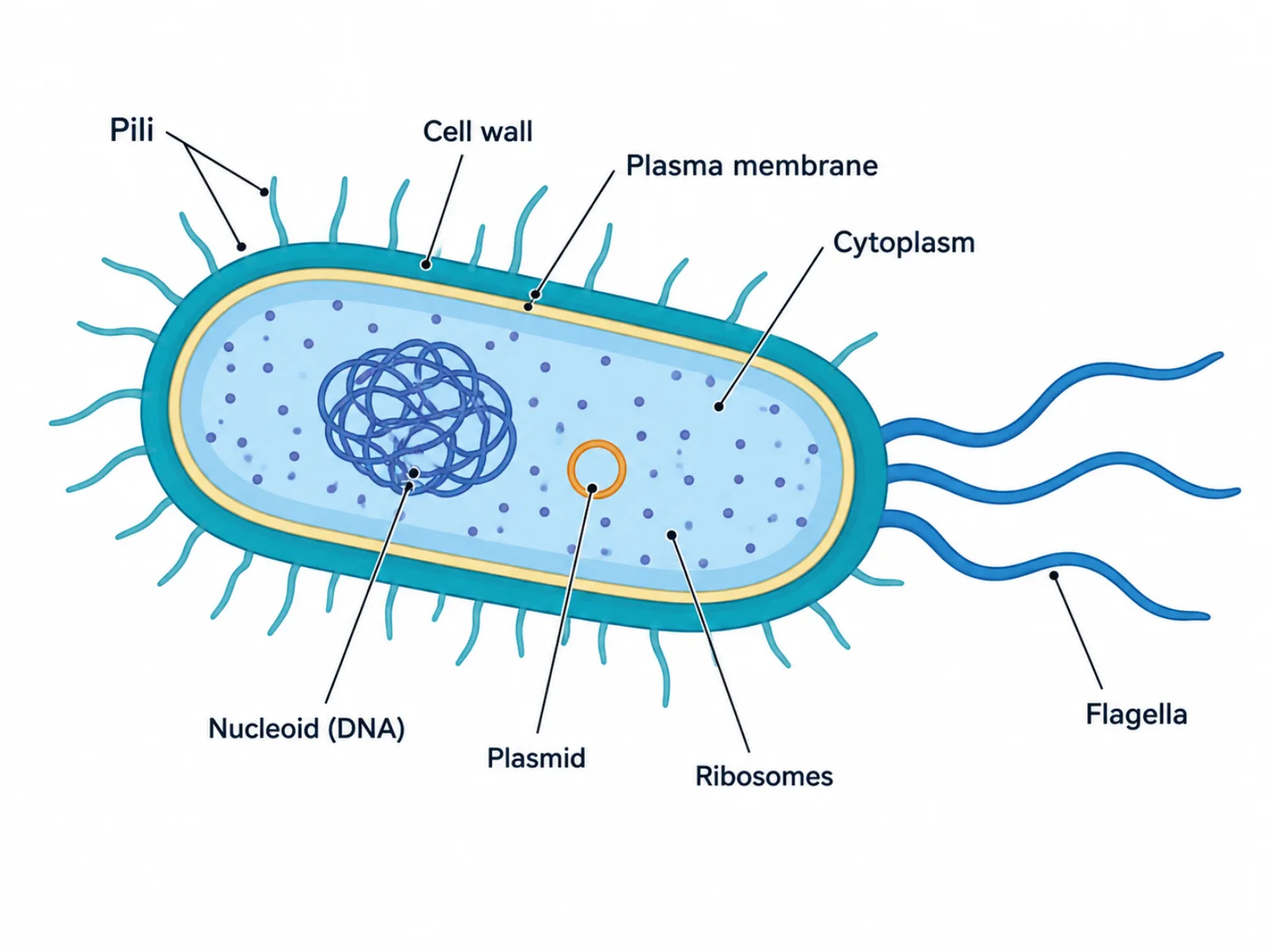
At a glance, a prokaryotic cell diagram shows a single enclosed compartment bounded by a cell wall and plasma membrane, with all internal contents sharing one open cytoplasmic space. Unlike diagrams of more complex cells, there are no internal membranes dividing the interior into separate rooms. This single-compartment design reflects real biology: prokaryotes are generally smaller and structurally simpler than eukaryotic cells, and their DNA, ribosomes, and metabolic machinery all operate in direct contact with one another rather than being sorted into dedicated organelles.
The Labeled Parts of a Prokaryotic Cell Diagram
- Cell wall — a rigid outer layer, made of peptidoglycan in bacteria, that gives the cell shape and protects it from bursting.
- Plasma membrane — the selectively permeable membrane just inside the cell wall, controlling what enters and exits the cell.
- Cytoplasm — the fluid interior where metabolic reactions, protein synthesis, and DNA all take place without separating membranes.
- Nucleoid — a region of cytoplasm holding the single circular chromosome, distinct from a nucleus because it has no enclosing membrane.
- Plasmid — a small circular piece of DNA separate from the main chromosome, often carrying genes such as antibiotic resistance.
- Ribosomes — smaller 70S ribosomes scattered throughout the cytoplasm, responsible for translating RNA into protein.
- Flagella — whip-like structures that rotate to propel the cell through liquid environments.
- Pili (fimbriae) — hair-like surface structures used for attaching to surfaces and for transferring DNA between cells during conjugation.
How to Interpret the Prokaryotic Cell Diagram: Reading a Cell Without a Nucleus
Read a prokaryotic cell diagram from the outside in: start at the cell wall, move through the plasma membrane, and land in the single open cytoplasmic space that contains everything else. Because there are no internal membranes, there is no strict "flow" or sequence the way there is in a process diagram — instead, the layout emphasizes proximity and scale. The nucleoid typically occupies a large, irregular area near the center, signaling that the chromosome is not confined but simply concentrated there, while one or more small circular plasmids are drawn separately to show they are physically distinct DNA molecules. Ribosomes are usually scattered as small dots throughout the cytoplasm rather than clustered, reflecting that translation happens simultaneously with transcription, since there is no nuclear envelope to separate the two processes in time or space.
Common Mistakes When Reading or Drawing a Prokaryotic Cell Diagram
The single most common error is drawing a nucleus — prokaryotic cells have no membrane-bound nucleus of any kind, only the unenclosed nucleoid region, and adding a nuclear envelope misrepresents the entire cell type. A closely related mistake is treating "nucleoid" and "nucleus" as interchangeable terms; the nucleoid is a region within an open cytoplasm, while a nucleus is a discrete organelle bounded by its own double membrane. A third common error is forgetting that the plasmid is a second, separate piece of DNA distinct from the main chromosome — some diagrams omit it entirely or merge it visually with the nucleoid, which obscures the fact that plasmids can be gained, lost, or exchanged independently of the chromosome itself.
Prokaryotic vs. Eukaryotic Cell Diagrams
The clearest way to understand a prokaryotic cell diagram is to contrast it directly with a eukaryotic cell diagram. Prokaryotic cells have no nucleus and no membrane-bound organelles at all; their DNA exists as a single circular chromosome floating in the cytoplasm, and they are generally much smaller than eukaryotic cells. Eukaryotic cells, by comparison, compartmentalize their genetic material inside a true membrane-bound nucleus and rely on dedicated organelles — mitochondria, the endoplasmic reticulum, the Golgi apparatus — to divide labor across the cell. Every structure that a prokaryotic cell diagram omits (nucleus, mitochondria, internal membrane systems) is precisely what distinguishes it from its eukaryotic counterpart, making this comparison one of the fastest ways to check whether a diagram has been drawn correctly.