
Neuron Diagram: Labeled Parts and Signal Pathway
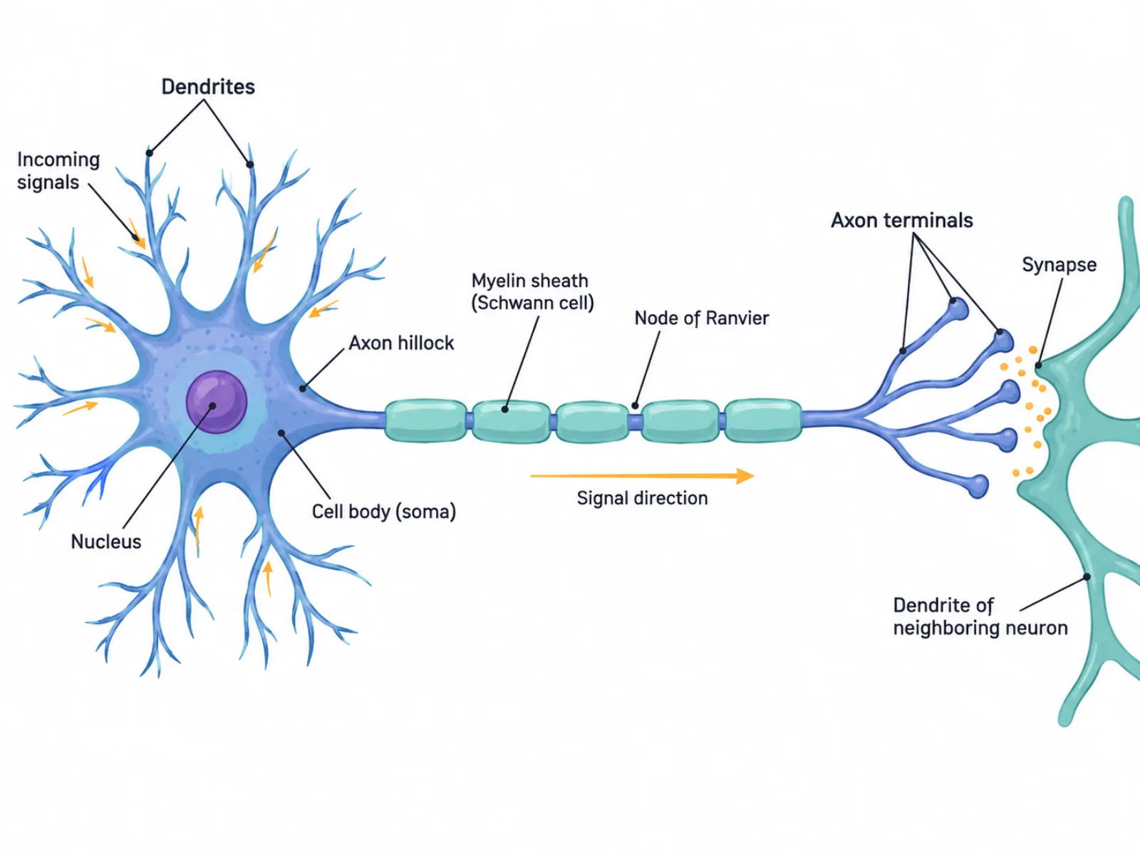
A neuron diagram is a labeled illustration of a nerve cell that shows both its physical structures and the path an electrical signal takes as it moves through the cell. Because neurons are elongated and asymmetric — unlike the roughly round cells shown in most biology textbooks — a neuron diagram has to stretch across the page to capture the full distance from the receiving end to the sending end. Reading one correctly means understanding that the diagram is not just a static map of parts; it's also a flowchart of signal direction, and the two purposes are drawn onto the same figure.
What a Neuron Diagram Shows
The diagram represents a single nerve cell specialized for receiving, integrating, and transmitting electrical impulses. Unlike most cells, which are compact and roughly symmetrical, a neuron is stretched into a distinct shape: a branching receiving end, a rounded cell body, and a long conducting fiber ending in a signal-releasing tip. The layout of a neuron diagram reflects this division of labor — each region of the cell has a specific job in moving information from one neuron to the next, and the diagram's left-to-right or top-to-bottom arrangement usually mirrors the actual direction the signal travels.
The Labeled Parts of a Neuron Diagram
- Dendrites — short, branching extensions that receive incoming signals from other neurons.
- Soma (cell body) with nucleus — integrates the incoming signals from the dendrites and houses the cell's genetic material and main metabolic machinery.
- Axon hillock — the trigger zone at the base of the axon where summed signals must reach a threshold to initiate an action potential.
- Axon — the single long fiber that conducts the electrical impulse away from the cell body toward the next neuron.
- Myelin sheath (formed by Schwann cells) — a fatty insulating layer wrapped around the axon in segments, which speeds up signal conduction.
- Nodes of Ranvier — small gaps between myelin segments where the axon membrane is exposed, allowing the impulse to "jump" from node to node in saltatory conduction.
- Axon terminals (synaptic boutons) — the branched endings of the axon that release neurotransmitter chemicals.
- Synapse — the junction between an axon terminal and the dendrite of the next neuron, where the signal is chemically transmitted across a small gap.
How to Interpret the Diagram: Tracing Signal Direction
The clearest way to read a neuron diagram is to follow the signal's path in strict order: dendrites receive input, the soma integrates it, the axon hillock decides whether to fire, the axon conducts the impulse, and the axon terminals release neurotransmitter into the synapse. This is a one-way path — information does not flow backward through the same neuron. Diagrams often use arrows along the axon to reinforce this direction, and the myelin sheath is typically drawn as a series of separate segments rather than one unbroken tube, precisely because the nodes of Ranvier between segments are functionally important, not just decorative gaps. When drawing a neuron diagram, keep the axon proportionally long relative to the cell body and dendrites, since in real neurons the axon can extend far beyond the compact cluster of dendrites — this proportion is part of what makes the diagram accurate, not just legible.
Common Mistakes in Reading or Drawing a Neuron Diagram
The most common mistake is reversing the signal direction, for example assuming information flows from the axon terminals back toward the dendrites. Signal flow is strictly one-directional within a single neuron: dendrite to soma to axon to terminal. A second frequent error is confusing dendrites with the axon, since both are thin projections from the cell body; the distinguishing features are that dendrites are short, numerous, and branch close to the soma to receive signals, while the axon is typically a single long fiber dedicated to sending the signal onward. A third mistake is drawing or imagining the myelin sheath as one continuous, unbroken coating along the entire axon. In reality, myelin is deposited in discrete segments by Schwann cells, with exposed nodes of Ranvier in between — this segmented structure is what enables the rapid, hopping conduction that makes myelinated neurons so much faster than unmyelinated ones.
Neuron Diagram vs. Generic Animal Cell Diagram
A neuron diagram is best understood as a specialized version of a generic animal cell diagram rather than a completely different kind of figure. Both include a nucleus, mitochondria, and other standard organelles needed to keep the cell alive and metabolically active. What sets the neuron diagram apart is the addition of structures built specifically for communication: the branching dendrites, the elongated axon wrapped in a myelin sheath, and the synaptic terminals at the end. A generic animal cell diagram typically shows a round or irregular cell with organelles distributed fairly evenly around a central nucleus, since its purpose is to illustrate general cell biology. A neuron diagram, by contrast, stretches that same basic cell body into an extreme, elongated shape because its entire structure is organized around a single function — moving an electrical signal as quickly and reliably as possible from one end of the cell to the other, sometimes across a distance of more than a meter in the human body.