
Human Eye Diagram: Labeled Parts and Light Path
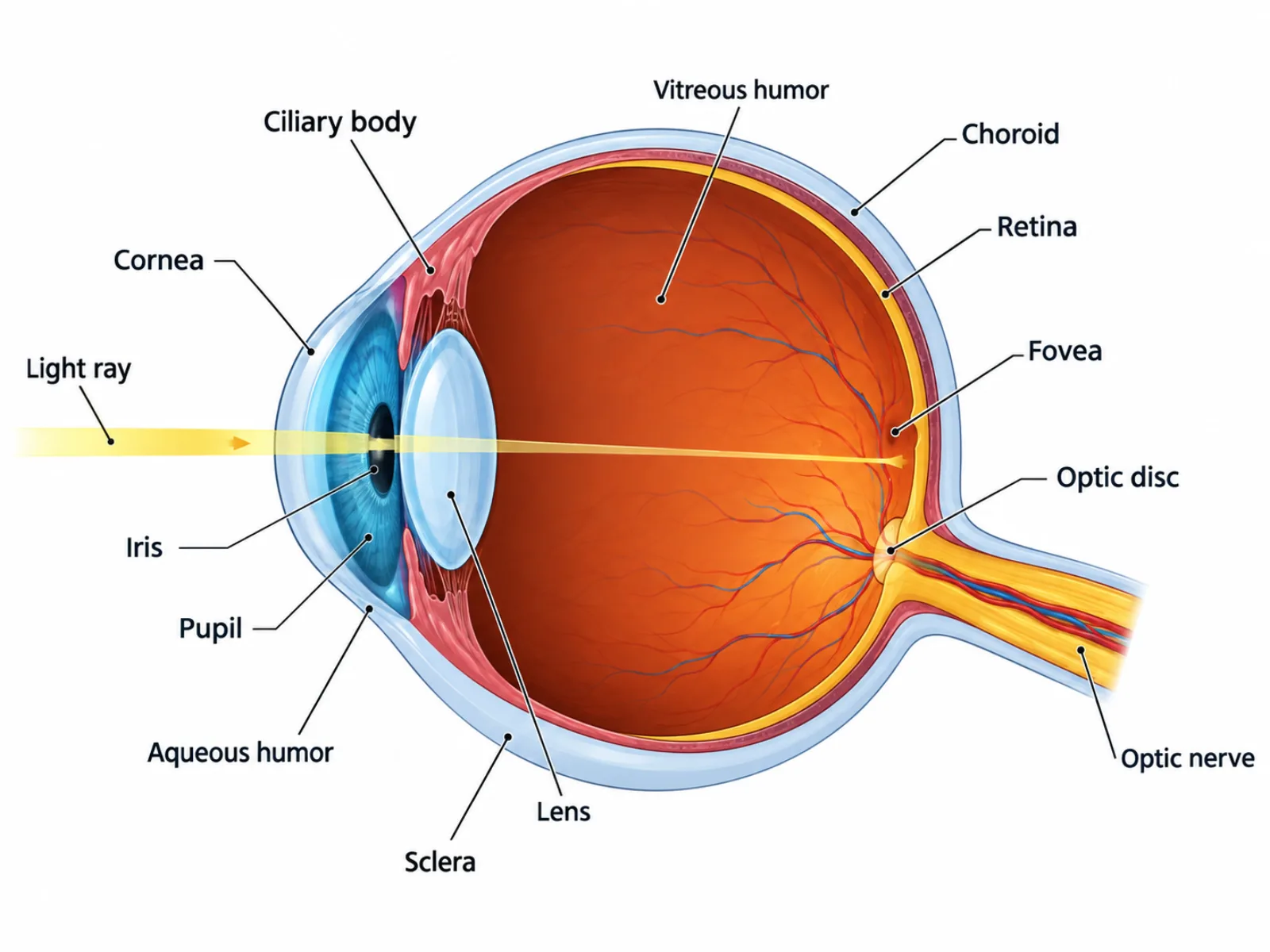
A human eye diagram is a cross-sectional illustration that shows how light entering the eye is bent, focused, and converted into the electrical signals the brain interprets as vision. Rather than depicting the eye as a solid sphere, a labeled human eye diagram slices it open to reveal the layered structures — cornea, lens, retina, and the fluid-filled chambers between them — arranged along the path that light actually travels. Because so many of these structures look similar in cross-section, a good human eye diagram depends on clear labeling and on tracing the light path in the correct order from front to back.
What a Human Eye Diagram Shows
At the broadest level, a human eye diagram represents an optical system built around a single job: bending incoming light so it converges precisely on a layer of light-sensitive cells at the back of the eye. The front portion of the diagram — cornea, iris, pupil, and lens — handles the physics of bending and regulating light. The back portion — retina, fovea, and optic nerve — handles converting that focused light into a signal the brain can use. A diagram that separates these two halves clearly makes it much easier to understand why damage to the front (like a clouded cornea) causes different symptoms than damage to the back (like retinal detachment).
The Labeled Parts of a Human Eye Diagram
- Cornea — the transparent front layer of the eye that performs most of the light bending, or refraction, before light ever reaches the lens.
- Iris — the pigmented muscular ring that gives the eye its color and controls the size of the pupil in response to light levels.
- Pupil — the opening at the center of the iris through which light enters the eye.
- Lens — a flexible structure behind the iris that fine-tunes focus by changing shape, a process called accommodation.
- Ciliary body and ciliary muscles — attached to the lens by zonule fibers, these muscles contract or relax to reshape the lens for near or distant focus.
- Aqueous humor — the watery fluid filling the front chamber between the cornea and lens, maintaining eye pressure and nourishing nearby tissue.
- Vitreous humor — the thicker, gel-like fluid filling the large chamber behind the lens, helping the eyeball hold its spherical shape.
- Retina — the light-sensitive layer lining the back of the eye, containing photoreceptors called rods and cones that convert light into electrical signals.
- Fovea — a small pit within the retina densely packed with cones, marking the point of sharpest central vision.
- Optic nerve and optic disc — the nerve that carries visual signals to the brain; the optic disc, where it exits the eye, has no photoreceptors and creates the blind spot.
- Sclera — the tough, white, fibrous outer coat that gives the eyeball its shape and protects the internal structures.
- Choroid — a vascular layer between the sclera and retina that supplies blood to the eye and absorbs stray light to reduce internal glare.
How to Read the Diagram: Tracing the Path of Light
The correct way to interpret a human eye diagram is to follow the path of light from the outside in, rather than reading the labels in whatever order they appear on the page. Light first strikes the cornea, which does the bulk of the refractive bending; it then passes through the aqueous humor, the pupil, and the lens, which makes final adjustments to focus the image precisely. From there, light crosses the vitreous humor and lands on the retina, where the rods and cones convert it into electrical impulses. Those impulses travel along the optic nerve to the brain. Proportions matter in this diagram too — the cornea and lens together occupy only a small fraction of the eye's total volume, while the vitreous chamber behind the lens takes up most of the eyeball's interior, which is why the eye holds its round shape even under pressure.
Common Mistakes When Reading a Human Eye Diagram
One of the most frequent errors is assuming the retina itself produces an upright image; in reality, the cornea and lens invert the image as they focus it, and the retina receives it upside down — the brain is what reconstructs an upright perception from that inverted signal. A second common mistake is confusing the optic disc with the fovea, since both appear as small distinct points on the retina in most diagrams; the two have essentially opposite roles, as the optic disc entirely lacks photoreceptors and creates the blind spot, while the fovea has the eye's densest concentration of cones and produces the sharpest vision. A third mistake is misordering the light path, such as placing the lens before the cornea or forgetting that light must cross both the aqueous and vitreous humor before reaching the retina.
How a Human Eye Diagram Differs from a Neuron Diagram
It helps to see the human eye diagram in relation to a neuron diagram, since the two are closely connected but represent different scales of biology. The retina's rods and cones are themselves specialized neurons, meaning the eye diagram is really an organ-level illustration built around a chain of neural signal conversion — light energy becomes an electrical impulse that then travels along the optic nerve. A neuron diagram, by contrast, zooms in to isolate the structure of a single cell, showing components like the dendrites, cell body, axon, and synaptic terminals that apply to any neuron in the body, not just those in the eye. In short, the eye diagram shows an entire optical organ built from many specialized neurons and supporting tissues working together, while the neuron diagram shows the anatomy of just one of those cells in isolation.