
Respiratory System Diagram: Labeled Gas Exchange
A respiratory system diagram is an anatomical illustration that traces the pathway air takes from the nose or mouth down through a branching network of tubes into the lungs, ending at the microscopic sacs where oxygen finally enters the bloodstream. Unlike a single-organ diagram, a respiratory system diagram has to represent a whole pathway, since air passes through several distinct structures — each with cartilage, muscle, or tissue suited to its specific job — before gas exchange can occur. Reading it correctly means understanding not just what each structure is called, but why the pathway narrows and branches the way it does on its way to the lungs.
What a Respiratory System Diagram Shows
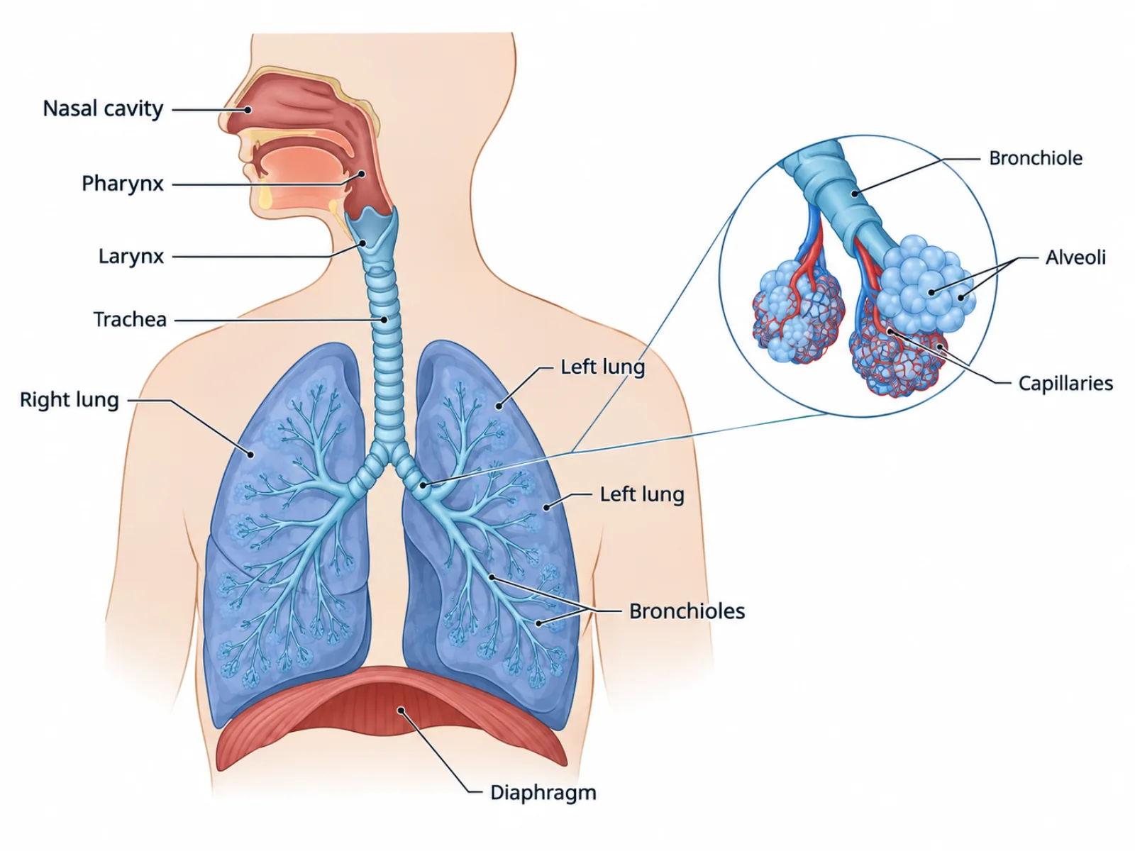
At its core, a respiratory system diagram represents a single continuous airway that starts wide and public — shared with the digestive tract at the top — and ends narrow and private, dedicated entirely to gas exchange. The upper portion of the diagram conditions incoming air, filtering and warming it before it goes any deeper. The middle portion is essentially plumbing: a branching tree of tubes that gets smaller and more numerous the further it goes. The lower portion is where the actual biological work happens, at a scale so small it can only be shown as a magnified inset in most diagrams. Recognizing this progression — from conditioning to conducting to exchanging — is the fastest way to make sense of the whole layout.
The Labeled Parts of a Respiratory System Diagram
- Nasal cavity — filters, warms, and humidifies air as it first enters the body.
- Pharynx — a shared passage behind the nasal cavity and mouth used by both the respiratory and digestive tracts.
- Larynx (voice box) — houses the vocal cords for sound production; its epiglottis folds down during swallowing to keep food out of the airway.
- Trachea (windpipe) — the main airway leading to the lungs, held permanently open by C-shaped rings of cartilage.
- Bronchi — the two main branches of the trachea, one entering each lung, still reinforced by cartilage like the trachea.
- Bronchioles — progressively smaller tubes branching from the bronchi, using smooth muscle rather than cartilage to control airflow.
- Alveoli — tiny thin-walled air sacs clustered at the ends of the bronchioles, surrounded by capillaries; this is the actual site of gas exchange.
- Diaphragm — a dome-shaped muscle below the lungs that contracts and flattens to expand the chest cavity and pull air in.
How to Read the Diagram: Following the Airway from Top to Bottom
The most reliable way to interpret a respiratory system diagram is to trace the path of a single breath from the nose down to the alveoli, following the branching structure rather than jumping between labels. Air enters through the nasal cavity, passes through the pharynx and larynx, and travels down the trachea, which splits into the two bronchi at the lungs. Each bronchus then divides repeatedly into smaller and smaller bronchioles, a branching pattern that resembles an upside-down tree, until the airway terminates in clusters of alveoli. Proportions in the diagram matter here: the trachea and bronchi are wide and relatively few in number, while the bronchioles and alveoli are extremely numerous and small, since the total surface area for gas exchange depends on having millions of tiny alveoli rather than a few large ones. The diaphragm is usually drawn beneath the lungs as a separate muscular structure, and its contraction and relaxation — not the lungs pushing themselves — is what actually drives the physical process of inhaling and exhaling.
Common Mistakes When Reading or Drawing a Respiratory System Diagram
The most frequent error is confusing bronchi with bronchioles, since both are branching airway tubes that look similar in a simplified diagram; the distinguishing feature is that bronchi are supported by cartilage rings like the trachea, while bronchioles rely on smooth muscle instead. A second common mistake is assuming that gas exchange happens somewhere in the bronchioles, when it is actually restricted to the alveoli at the very end of the branching network, where thin walls and a dense capillary network allow oxygen and carbon dioxide to diffuse across. A third mistake is leaving out or misplacing the diaphragm, treating breathing as something the lungs do on their own rather than recognizing that the diaphragm's mechanical contraction is what creates the pressure difference driving air in and out.
Respiratory System vs. Cellular Respiration: A Frequently Confused Pair
Because both terms share the word "respiration," students very often conflate a respiratory system diagram with a cellular respiration diagram, even though they describe entirely different levels of biology. A respiratory system diagram shows the physical passageway — nose, trachea, bronchi, bronchioles, and alveoli — that brings oxygen into the blood and removes carbon dioxide at the level of whole organs. Cellular respiration, by contrast, is a biochemical process that takes place inside individual cells, specifically in the mitochondria, where that oxygen delivered by the lungs is actually used to break down glucose and produce ATP, the cell's usable energy currency. One diagram is anatomy: tubes, muscles, and air sacs you could point to in a dissected body. The other is metabolism: a sequence of chemical reactions you would only see represented as arrows and molecule names. Keeping this distinction clear prevents a common exam mistake of describing oxygen "being used for energy" at the alveoli, when the alveoli only handle the physical exchange of gases with the blood — the actual energy-producing chemistry happens later, inside cells throughout the body.