
DNA Replication Diagram: Fork and Strand Synthesis
A DNA replication diagram shows how a cell copies its genetic material before dividing, capturing the double helix in the act of being unwound and rebuilt rather than sitting at rest. Instead of a static ladder, a DNA replication diagram depicts an open, Y-shaped fork where enzymes work on both separated strands at once, producing two new strands by different mechanisms because the original strands run in opposite directions. Understanding this diagram means following each enzyme's specific job in order, from the first opening of the helix to the final sealed strand.
What the DNA Replication Diagram Shows
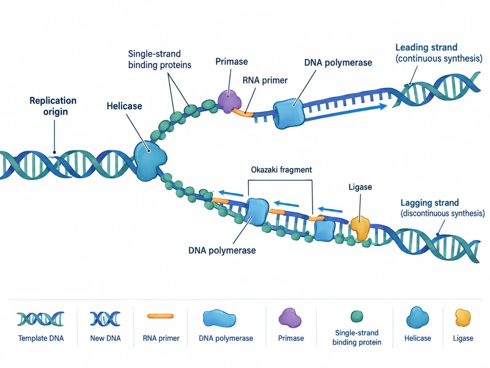
The diagram captures a moment mid-process: the double helix has already begun to unwind, exposing two single strands that each serve as a template for a new complementary strand. Because both new strands are built from the same fork at the same time but the templates run antiparallel to each other, the diagram has to show two different synthesis styles happening side by side — one smooth and continuous, one broken into pieces. This asymmetry is the single most important idea a DNA replication diagram communicates, and everything else in the image exists to explain why it happens.
The Labeled Parts of a DNA Replication Diagram
- Origin of replication — the specific site on the DNA molecule where the helix first opens, giving replication machinery a starting point.
- Helicase — an enzyme that unwinds the double helix by breaking the hydrogen bonds holding the two strands together, moving the fork forward.
- Single-strand binding proteins (SSBs) — coat the newly separated strands to keep them from snapping back together (re-annealing) before they can be copied.
- Primase — synthesizes a short RNA primer, giving DNA polymerase the free 3' end it needs to begin adding nucleotides.
- DNA polymerase — builds new DNA by adding nucleotides only in the 5'-to-3' direction, and cannot start a strand without a primer already in place.
- Leading strand — synthesized continuously in one smooth piece because its template allows polymerase to move in the same direction the fork is opening.
- Lagging strand — synthesized discontinuously, in short segments called Okazaki fragments, because its template runs the opposite direction from fork movement.
- DNA ligase — seals the gaps between adjacent Okazaki fragments, joining them into one unbroken lagging strand.
- Replication fork — the Y-shaped junction where the helix is actively unwinding and both new strands are being assembled.
How to Read the Diagram
Read a DNA replication diagram by following the fork's direction of travel first, then tracking each template strand separately. Start at the origin of replication, where helicase has already pried the helix open, and note that SSBs line both exposed strands to hold them apart. On the strand that runs so polymerase can follow the fork directly, synthesis proceeds as one continuous line — this is the leading strand. On the other template, polymerase must repeatedly restart, laying a primer, extending a short fragment, then jumping back toward the fork to start again — these fragments are what give the lagging strand its stitched-together look, until ligase joins them. Arrows in the diagram typically point in the 5'-to-3' direction of the newly synthesized strand, not the template, so tracing arrow direction tells you which strand is being built and which way polymerase is moving relative to the fork.
Common Mistakes When Reading or Drawing a DNA Replication Diagram
A frequent error is assuming the lagging strand is synthesized more slowly or less accurately than the leading strand — in reality both are built at comparable speed and fidelity; the only difference is continuity, driven purely by the antiparallel orientation of the two template strands. Another common mistake is drawing or assuming DNA polymerase can initiate a brand-new strand without a primer; polymerase can only extend an existing 3' end, which is exactly why primase must act first on both strands. Students also frequently confuse helicase and ligase because both act directly on the DNA backbone: helicase opens the helix ahead of the fork, while ligase closes gaps behind it — one separates, the other joins.
DNA Replication Diagram vs. DNA Structure Diagram
A DNA structure diagram is a static picture: it shows the double helix at rest, emphasizing base pairing, the sugar-phosphate backbone, and the helix's twisted geometry, with no fork and no enzymes in motion. A DNA replication diagram, by contrast, is a process diagram — it freezes a dynamic event mid-way, showing the helix partially opened, enzymes actively bound to the strands, and two new strands under construction by different mechanisms. Where a structure diagram answers "what does DNA look like," a replication diagram answers "how does a cell make a second copy of it." This copying event is not a standalone process; it takes place during the S phase of the cell cycle, completing before the cell proceeds into mitosis, where the two identical copies are distributed to daughter cells.