
DNA Structure Diagram: Double Helix and Base Pairs
A DNA structure diagram is a labeled illustration of the double helix — the twisted, ladder-like arrangement of two nucleic acid strands held together by paired bases. Because DNA is a molecule, not a visible object, this kind of diagram is the primary way students and researchers actually see its architecture: the two backbones spiraling around each other, the bases stacked like rungs between them, and the grooves left exposed along the outside. A well-drawn DNA structure diagram makes a set of otherwise abstract chemical rules — antiparallel strands, base-pairing specificity, hydrogen bonding — visually obvious at a glance.
What the DNA Structure Diagram Shows
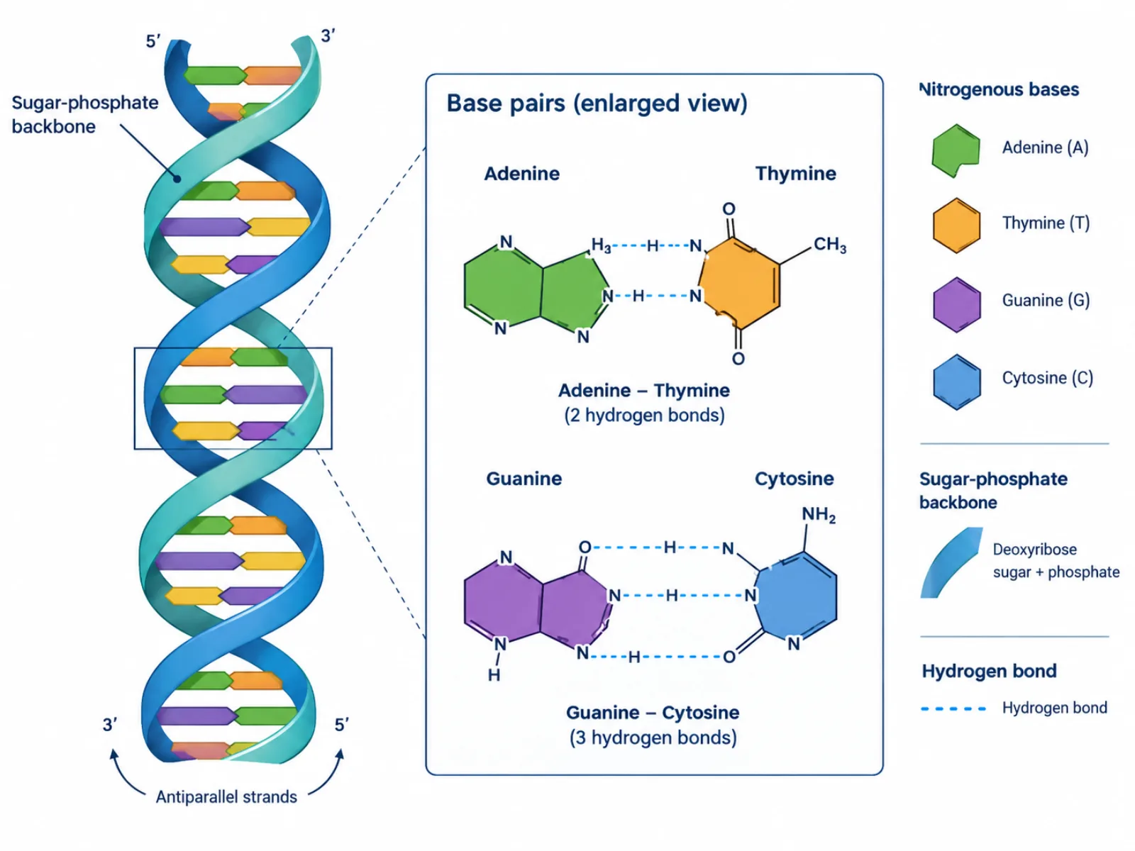
At its simplest, the diagram represents two long strands twisted around a shared axis, forming the double helix first described from X-ray diffraction data. Each strand is a chain of nucleotides, and the two chains are connected across the middle by pairs of nitrogenous bases. The diagram's job is to make three separate facts visible at once: the physical shape of the helix, the chemical identity of each backbone strand, and the specific pairing rules that hold the two sides together. A flat, two-dimensional "ladder" version of the diagram is often used for teaching base pairing, while a helical rendering is used to show the molecule's true three-dimensional geometry.
The Labeled Parts and What They Do
- Sugar-phosphate backbones — two continuous strands, each built from alternating deoxyribose sugar and phosphate groups, that form the structural rails of the helix and hold the genetic information's chemical framework together.
- Antiparallel orientation — the two backbones run in opposite 5'-to-3' directions relative to each other, which is a structural requirement for the bases to pair correctly and for replication enzymes to read the strands.
- Adenine (A) — a purine base with a double-ring structure that always pairs with thymine.
- Guanine (G) — a purine base with a double-ring structure that always pairs with cytosine.
- Thymine (T) — a pyrimidine base with a single-ring structure that always pairs with adenine via two hydrogen bonds.
- Cytosine (C) — a pyrimidine base with a single-ring structure that always pairs with guanine via three hydrogen bonds.
- Base pairs (rungs) — the paired bases spanning the center of the helix, held together by hydrogen bonds and stacked on top of one another to stabilize the structure.
- Major groove — the wider gap spiraling along the helix's exterior, wide enough for many DNA-binding proteins to insert and read the base sequence directly.
- Minor groove — the narrower gap on the opposite side of the same spiral, used by a different set of regulatory proteins and DNA-binding molecules.
How to Read and Draw a DNA Structure Diagram
Reading a DNA structure diagram correctly starts with orientation: identify which end of each backbone is the 5' end and which is the 3' end, since the two strands must run in opposite directions for the diagram to be accurate. From there, check every rung — each base pair should show one purine (A or G) opposite one pyrimidine (T or C), never two purines or two pyrimidines paired together, since a purine-purine pair would be too wide for the helix and a pyrimidine-pyrimidine pair too narrow. When drawing the diagram, the hydrogen bonds between A-T should be shown as two connecting lines and between G-C as three, reflecting the actual bond count and giving G-C pairs slightly greater chemical stability. The grooves are typically labeled last, as thin spiraling channels along the outside of the twisted backbones rather than part of the rungs themselves.
Common Mistakes to Avoid
The most frequent error is mispairing bases — putting adenine with guanine, or cytosine with thymine — which breaks the geometric fit of the helix and violates Chargaff's base-pairing rules entirely. A second common mistake is drawing both backbones running in the same direction; DNA's two strands are always antiparallel, and this detail matters because replication and transcription enzymes rely on that opposite orientation to move along the molecule correctly. A third mistake is conflating a "nucleotide" with a "base": a nucleotide is the full repeating unit of sugar, phosphate, and base together, while the base is only the nitrogen-containing ring portion that does the pairing. Labeling a full nucleotide as if it were just a base — or vice versa — misrepresents what the backbone is actually made of.
DNA Structure vs. DNA Replication Diagrams
A DNA structure diagram and a DNA replication diagram are often confused because they depict the same molecule, but they capture different moments. The structure diagram shows DNA at rest — a stable, closed double helix with its bases already paired and its backbones intact — used to teach the molecule's static geometry and pairing rules. A DNA replication diagram instead shows the helix being actively unwound by enzymes, with the two original strands separated and new complementary strands being synthesized alongside each one. It's worth also noting that the double helix shown in a structure diagram is rarely DNA's final packaged form inside a living cell: in eukaryotes, the helix is further wound around histone proteins into chromatin, and then condensed further into chromosomes, which a basic structure diagram does not attempt to show.