
Chloroplast Diagram: Labeled Structure and Function
A chloroplast diagram is a cross-sectional illustration of the organelle in plant and algal cells responsible for photosynthesis, showing how its internal membranes are organized to capture light and convert it into chemical energy. Because photosynthesis happens in two distinct stages that occur in two distinct locations inside the organelle, a labeled chloroplast diagram has to clearly separate the membrane-bound compartments from the surrounding fluid, or the two stages of photosynthesis become impossible to tell apart. Reading the diagram accurately means understanding not just the names of the structures but which specific reactions happen inside each one.
What a Chloroplast Diagram Shows
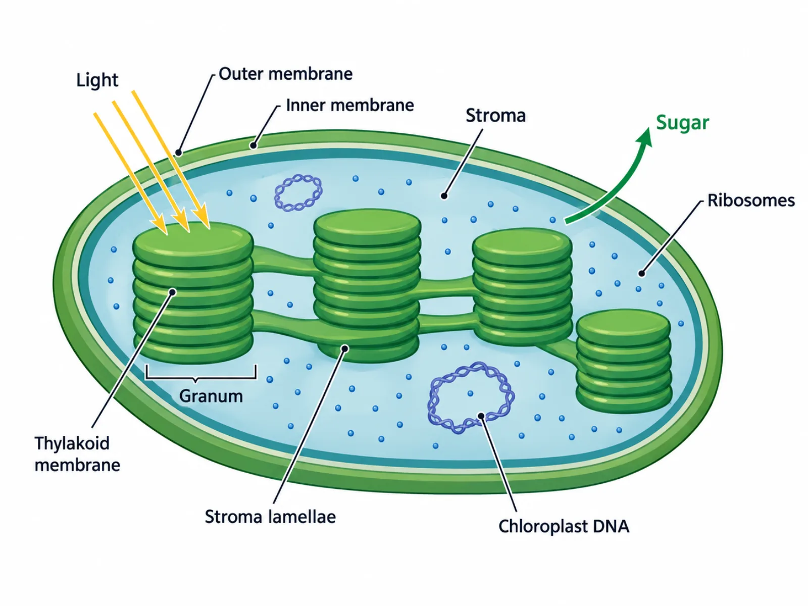
At a basic level, a chloroplast diagram represents an organelle organized around a central strategy: separating the light-capturing machinery from the sugar-building machinery so each can operate under its own conditions. The outer boundary consists of two membranes enclosing a fluid interior called the stroma. Suspended within that stroma is a separate internal membrane system, folded into flattened sacs and stacked into columns. This nested structure — membranes within membranes — is what allows a single organelle to run two chemically different processes side by side without them interfering with each other.
The Labeled Parts of a Chloroplast Diagram
- Outer membrane and inner membrane — chloroplasts are double-membrane organelles, with these two membranes enclosing the entire internal structure.
- Stroma — the fluid-filled interior of the chloroplast, where the Calvin cycle (the light-independent reactions that build sugar) takes place.
- Thylakoid — a single flattened, membrane-bound sac where the light-dependent reactions occur and where chlorophyll and other light-absorbing pigments are embedded.
- Granum (plural: grana) — a stack of thylakoids, resembling a stack of coins, formed to maximize the surface area available for capturing light.
- Stroma lamellae — thin connecting membranes that link separate grana together into one continuous network.
- Chloroplast DNA and ribosomes — chloroplasts carry their own small circular genome and their own ribosomes, evidence that they originated as free-living bacteria.
How to Read the Diagram: Two Reactions, Two Locations
The key to interpreting a chloroplast diagram correctly is to map each stage of photosynthesis to its specific location rather than treating the whole organelle as one undifferentiated space. Light strikes the thylakoid membrane first, where embedded pigments absorb its energy and use it to produce energy-carrying molecules; this is the light-dependent stage, and it is confined entirely to the thylakoid membrane inside the grana. Those energy-carrying molecules then diffuse into the surrounding stroma, where they power the Calvin cycle, the light-independent stage that actually assembles carbon dioxide into sugar. A diagram usually shows this relationship with the grana drawn as stacked discs suspended within the larger stroma space, and the proportion matters: a chloroplast typically contains many grana connected by stroma lamellae, maximizing the membrane surface area available for light capture relative to the surrounding stroma volume.
Common Mistakes When Reading or Drawing a Chloroplast Diagram
The most frequent mistake is confusing "granum" with "thylakoid" — a thylakoid is one single flattened sac, while a granum is the entire stack of thylakoids piled on top of each other, so calling one thylakoid a granum understates the structure it actually describes. A second common mistake is placing the light-dependent reactions in the stroma instead of the thylakoid membrane; the Calvin cycle does happen in the stroma, but light capture and the reactions it drives are strictly confined to the thylakoid membrane where the pigments are embedded. A third mistake is forgetting that the chloroplast is bounded by two membranes rather than one, which matters because that double membrane is a structural feature directly tied to how chloroplasts are believed to have originated.
How a Chloroplast Differs from a Mitochondrion
A chloroplast diagram is frequently compared to a mitochondrion diagram, since the two organelles share several structural similarities despite doing essentially opposite jobs. Both are double-membrane organelles, and both carry their own small circular DNA and ribosomes separate from the cell's nucleus — a major piece of evidence for the endosymbiotic theory, which holds that both organelles were once independent bacteria engulfed by an ancestral cell and retained for their useful functions. Functionally, though, they run in opposite directions: a chloroplast captures light energy and uses it to build sugar molecules through photosynthesis, taking in carbon dioxide and releasing oxygen, while a mitochondrion takes those sugar molecules and breaks them down through cellular respiration to release usable energy as ATP, taking in oxygen and releasing carbon dioxide. In a sense, the two organelles form a cycle: chloroplasts build the fuel that mitochondria later spend, whether that split happens across different organisms or, in plant cells, within the very same cell that contains both.