
Eukaryotic Cell Diagram: Nucleus and Organelles
A eukaryotic cell diagram illustrates the internal architecture of cells that organize their contents into distinct, membrane-bound compartments, most notably a true nucleus. Unlike simpler cell types, a eukaryotic cell diagram shows genetic material sealed inside its own envelope and shows dedicated organelles handling energy production, protein processing, and structural support as separate, specialized units. Reading it correctly requires recognizing that "eukaryotic" is a broad category covering animal, plant, fungal, and protist cells alike, not a synonym for animal cells specifically.
What a Eukaryotic Cell Diagram Shows
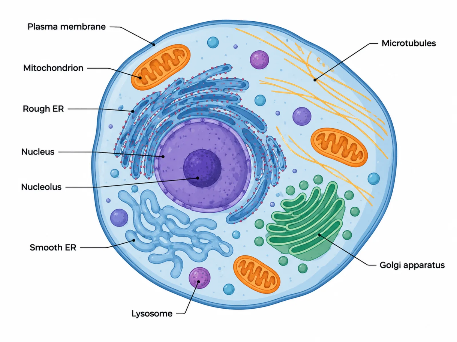
A eukaryotic cell diagram depicts a cell whose interior is subdivided by internal membranes into functionally distinct regions, in contrast to the single open cytoplasmic space of simpler cell types. This compartmentalization is the defining feature the diagram must convey: DNA sits inside a nucleus, energy production happens inside mitochondria, and protein processing occurs across a connected series of membrane-bound organelles. The overall message is one of division of labor — each compartment does one job well, rather than every process happening in one shared space.
The Labeled Parts of a Eukaryotic Cell Diagram
- Nucleus — a true membrane-bound organelle enclosing the cell's linear chromosomes; it houses the nucleolus, which assembles ribosomal subunits before they are exported to the cytoplasm.
- Nucleolus — a dense region inside the nucleus dedicated to assembling ribosomal subunits.
- Endoplasmic reticulum — part of the endomembrane system, continuous with the nuclear envelope, where proteins and lipids are synthesized and initially processed.
- Golgi apparatus — receives proteins from the endoplasmic reticulum, modifies them further, and packages them into vesicles for their final destinations.
- Lysosomes — membrane-bound sacs of digestive enzymes that break down waste, damaged organelles, and engulfed material.
- Transport vesicles — small membrane-bound sacs that shuttle material between the endoplasmic reticulum, Golgi apparatus, lysosomes, and the plasma membrane.
- Mitochondria — double-membrane organelles that generate ATP through cellular respiration and carry their own separate DNA.
- Cytoskeleton — a network of microtubules, microfilaments, and intermediate filaments that gives the cell shape, enables internal transport, and pulls chromosomes apart during cell division.
How to Interpret the Eukaryotic Cell Diagram: Reading the Internal Membranes
Read a eukaryotic cell diagram by tracing the endomembrane system as a single connected pathway rather than a set of unrelated parts: the nuclear envelope is continuous with the endoplasmic reticulum, which hands proteins off to the Golgi apparatus via transport vesicles, which in turn buds off vesicles heading toward lysosomes or the plasma membrane. This sequence — nucleus to ER to Golgi to final destination — is the functional "flow" the diagram encodes, even though the organelles themselves are static structures. Separately, note that mitochondria are drawn independently of this pathway, since they operate on their own membrane system entirely dedicated to energy production, and the cytoskeleton is drawn as a mesh spanning the whole cell rather than a single discrete organelle, reflecting its structural rather than biochemical role.
Common Mistakes When Reading or Drawing a Eukaryotic Cell Diagram
A frequent mistake is assuming "eukaryotic cell" means "animal cell" — plants, fungi, and protists are eukaryotes too, so a generic eukaryotic cell diagram should include only structures common to all of them, and adding plant-only features like a cell wall or chloroplast (or animal-only features like centrioles) without labeling the diagram as plant- or animal-specific creates a misleading, hybrid image. Another common error is forgetting that both plant and animal cells contain mitochondria; students sometimes assume only plant cells need an energy-producing organelle because they also have chloroplasts, when in fact chloroplasts are an addition on top of mitochondria, not a replacement for them. A third mistake is treating the endomembrane system's components as isolated organelles rather than one interconnected pathway, which obscures how a protein actually moves from synthesis to its final location.
Eukaryotic vs. Prokaryotic Cell Diagrams
The clearest contrast for a eukaryotic cell diagram is with a prokaryotic cell diagram. Eukaryotic cells enclose their DNA in a true, membrane-bound nucleus and organize multiple linear chromosomes, while prokaryotic cells keep a single circular chromosome in an open nucleoid region with no surrounding membrane. Eukaryotic cells are also generally larger and far more compartmentalized, relying on organelles like mitochondria and the endoplasmic reticulum that prokaryotic cells simply lack. Within the eukaryotic category itself, this diagram represents the general parent structure: a plant cell diagram and an animal cell diagram are both specialized versions of it, each adding structures — a cell wall and chloroplasts for plants, centrioles for animals — on top of the shared eukaryotic foundation shown here.