
Mitosis Diagram: Labeled Phases and Cytokinesis
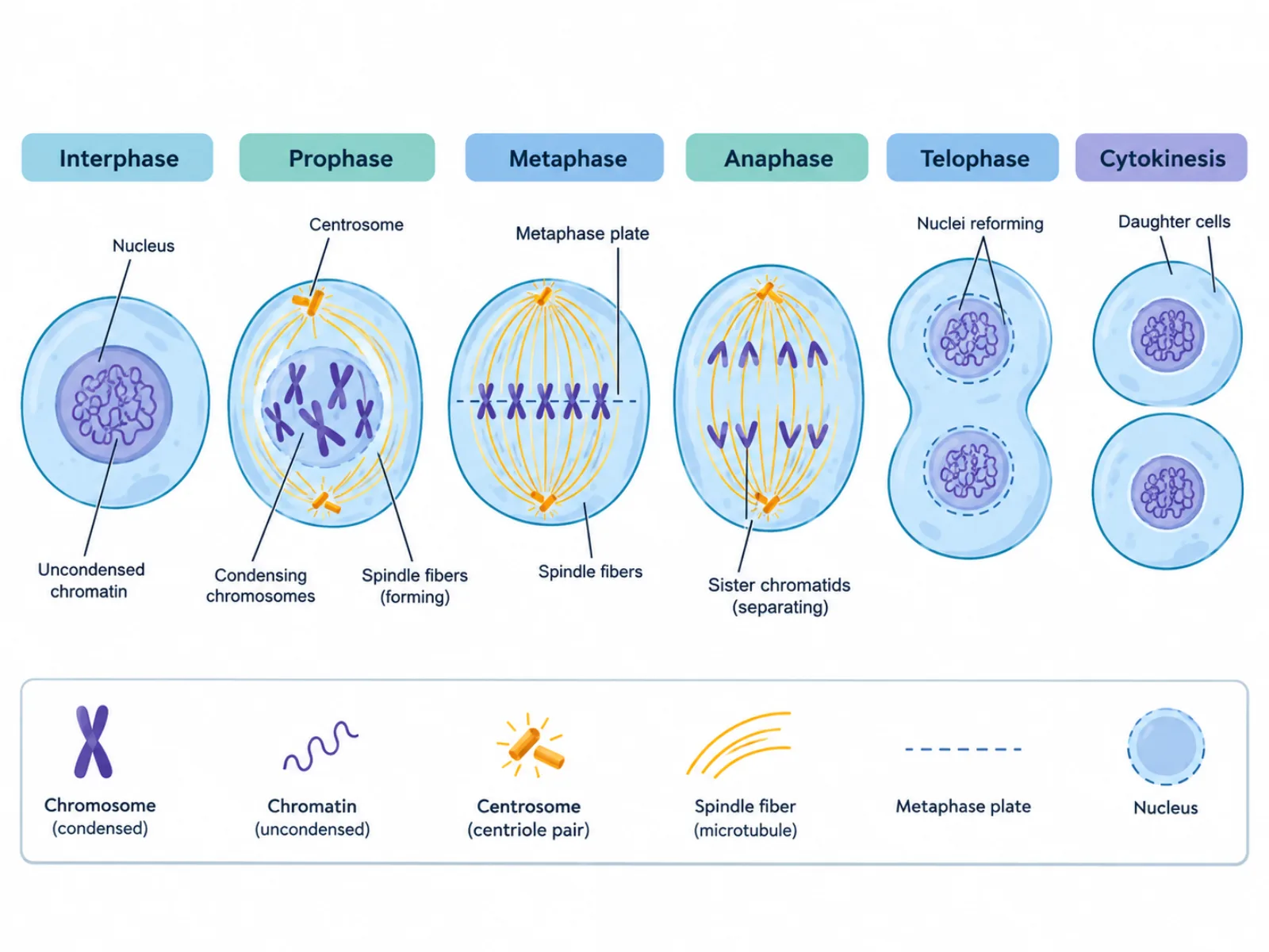
A mitosis diagram is a phase-by-phase illustration of how one cell divides into two genetically identical daughter cells, showing how chromosomes condense, align, separate, and are enclosed into two new nuclei. Because the phases of mitosis follow a strict order and each one looks visually distinct, a labeled mitosis diagram works best when it's read as a sequence rather than a collection of unrelated snapshots. Getting the order and the terminology right — especially the difference between a chromosome and a chromatid — is what separates a correct reading of the diagram from a common source of exam errors.
What a Mitosis Diagram Shows
At its core, a mitosis diagram represents the mechanism that allows a single cell to produce two functionally and genetically identical copies of itself, which is the basis for growth, tissue repair, and asexual reproduction in many organisms. The diagram typically starts with a cell that has already replicated its DNA and ends with two separate daughter cells, each containing a complete diploid set of chromosomes. Between those two points, the diagram breaks the process into distinct visual phases, each defined by what the chromosomes and the cell's internal structures are doing at that exact moment.
The Labeled Phases of a Mitosis Diagram
- Interphase — the cell grows and replicates its DNA before mitosis begins; technically not part of mitosis itself, but essential context for what the diagram shows next.
- Prophase — chromatin condenses into visible chromosomes, each made of two identical sister chromatids joined at a centromere; spindle fibers begin forming and the nuclear envelope breaks down.
- Metaphase — chromosomes line up individually along the metaphase plate at the cell's equator.
- Anaphase — sister chromatids are pulled apart by spindle fibers toward opposite poles of the cell.
- Telophase — chromosomes decondense and two new nuclear envelopes form around each separated set.
- Cytokinesis — the cytoplasm physically divides, producing two genetically identical diploid daughter cells.
How to Read the Diagram: Following the Phases in Order
The correct way to interpret a mitosis diagram is to read it strictly in phase order, since each stage is defined by a specific structural change that sets up the next one. Starting from interphase, where DNA replication has already doubled the genetic material, the diagram moves into prophase as chromosomes condense and become visible, then metaphase as those chromosomes align individually along the cell's center. Anaphase is the turning point of the whole sequence: this is the only phase where sister chromatids actually separate, and once they do, each chromatid is considered its own independent chromosome. Telophase and cytokinesis then close out the process, reforming nuclei and splitting the cytoplasm. Proportion and symmetry matter in this diagram: the two daughter cells produced at the end should be drawn as equal in size and identical in chromosome content, since that genetic identity is the entire point of mitotic division.
Common Mistakes When Reading or Drawing a Mitosis Diagram
The most common error is mixing up the terms "chromosome" and "chromatid" — a replicated chromosome with two sister chromatids still counts as one chromosome, not two, right up until anaphase, when the chromatids finally separate and each becomes its own chromosome. A second frequent mistake is confusing metaphase with anaphase, since both involve chromosomes positioned in the middle of the cell in simplified drawings; the distinguishing feature is that metaphase is about alignment (chromosomes lined up but not yet moving apart), while anaphase is about active separation (chromatids being pulled to opposite poles). A third mistake is treating cytokinesis as identical to telophase, when cytokinesis is a distinct final step involving the physical division of the cytoplasm, separate from the nuclear changes that define telophase.
How a Mitosis Diagram Differs from a Meiosis Diagram
A mitosis diagram is most usefully understood in contrast to a meiosis diagram, since the two describe different kinds of cell division with different biological purposes. Mitosis involves a single round of division, producing two daughter cells that are genetically identical to the parent cell and to each other, both retaining the original diploid chromosome number; there is no crossing over at any point in the process. Meiosis, by contrast, involves two consecutive rounds of division and produces four daughter cells, each haploid and genetically unique, thanks to crossing over during its first prophase and the independent assortment of chromosomes. Mitosis exists to support growth, tissue repair, and asexual reproduction, where genetic consistency is the goal; meiosis exists to produce gametes for sexual reproduction, where genetic variation is the goal. This single distinction — identical versus unique daughter cells, and two versus four of them — is usually the fastest way to tell which of the two diagrams you're looking at.