
Animal Cell Diagram: Labeled Organelles and Functions
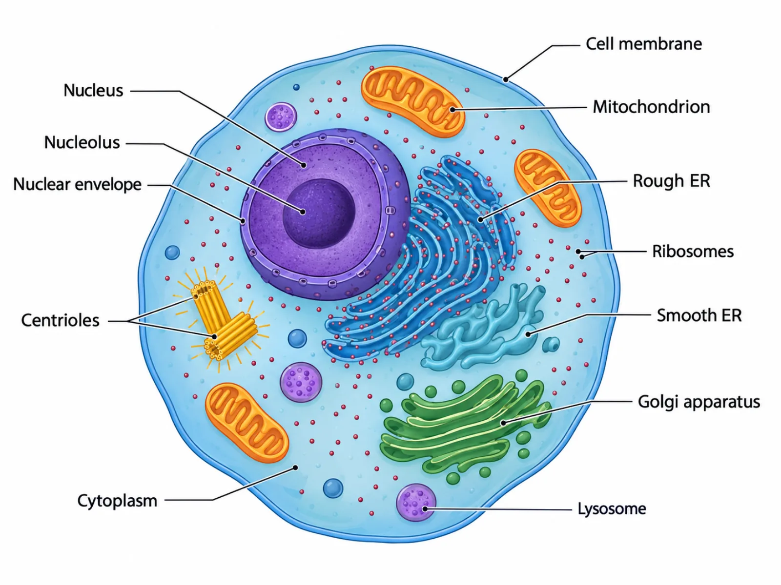
An animal cell diagram is a labeled cross-section of a typical animal cell, showing the organelles responsible for generating energy, synthesizing proteins, and maintaining the cell's internal organization. Because animal cells lack the rigid outer wall found in plants, an animal cell diagram is drawn with a rounded, often slightly irregular outline, and its organelles are distributed somewhat unevenly through a flexible interior rather than fitting neatly inside a fixed geometric shape. Reading an animal cell diagram well means recognizing not only what each labeled structure is, but also which structures are unique to animal cells and which are shared with virtually all eukaryotic cells.
What an Animal Cell Diagram Shows
The diagram represents the internal contents of a single animal cell as it would appear if sliced open and viewed from the side. Its purpose is to show how a cell that lacks rigid external support still maintains internal order, produces the energy it needs, and manufactures and processes the proteins and lipids necessary for life. Because animal cells can change shape, move, and divide in ways plant cells generally cannot, the diagram's flexible, rounded outline is itself informative — it signals that the cell's boundary is dynamic rather than fixed.
The Labeled Parts of an Animal Cell Diagram
- Cell membrane — a flexible barrier that controls what substances enter and exit the cell.
- Nucleus with nucleolus and nuclear envelope — houses the cell's DNA; the nucleolus within it manufactures ribosomal RNA, and the nuclear envelope separates the nucleus from the cytoplasm.
- Mitochondria — produce ATP through cellular respiration, supplying the energy the cell needs to function.
- Rough endoplasmic reticulum — studded with ribosomes, it carries out protein synthesis for molecules destined for secretion or membranes.
- Smooth endoplasmic reticulum — synthesizes lipids and assists in detoxifying certain compounds, lacking the ribosomes found on its rough counterpart.
- Golgi apparatus — modifies, sorts, and ships proteins to their correct destinations inside or outside the cell.
- Ribosomes — translate messenger RNA into protein, either freely in the cytoplasm or attached to the rough endoplasmic reticulum.
- Lysosomes — contain digestive enzymes that break down waste materials and worn-out organelles.
- Centrioles — organize the spindle fibers that separate chromosomes during cell division.
- Cytoplasm (cytosol) — the jelly-like medium filling the cell and suspending all of its organelles.
How to Interpret and Draw the Diagram
The most accurate way to read an animal cell diagram is to notice that, unlike a plant cell diagram, no single organelle dominates the interior space — organelles are distributed somewhat evenly throughout the cytoplasm, with the nucleus typically the largest single structure but rarely taking up more than a modest fraction of total volume. When drawing an animal cell diagram, the outline should be round or gently irregular rather than boxy, reflecting the absence of any rigid external wall. Mitochondria, ribosomes, and vesicles should be scattered rather than clustered in one spot, since these organelles perform their functions throughout the cytosol rather than in one fixed region. The nucleus is usually positioned centrally or slightly off-center, with the endoplasmic reticulum and Golgi apparatus arranged nearby, reflecting the real physical relationship between protein synthesis, processing, and packaging as one continuous production line inside the cell.
Common Mistakes When Reading or Drawing an Animal Cell Diagram
A frequent and significant error is adding a cell wall or chloroplasts to an animal cell diagram — both are exclusively plant structures and should never appear in an accurate animal cell illustration, since animal cells obtain energy by consuming food rather than photosynthesizing. Another common mistake is drawing only a single mitochondrion, which understates reality: most animal cells, particularly metabolically active ones like muscle or liver cells, contain hundreds to thousands of mitochondria distributed throughout the cytoplasm, not just one. A third mistake is giving the cell a fixed, rectangular, or otherwise geometrically regular outline; because there is no cell wall to enforce a rigid shape, real animal cells and accurate diagrams of them show a rounded or irregular boundary that can vary between cells and even change over time.
Animal Cell Diagram vs. Plant Cell Diagram
The clearest way to understand an animal cell diagram is by direct contrast with a plant cell diagram. An animal cell has no cell wall, only the flexible cell membrane, which is why it is drawn with a round or irregular outline rather than the fixed, boxy shape of a plant cell. An animal cell lacks a large central vacuole; instead it contains only small vesicles scattered through the cytoplasm, none large enough to dominate the cell's volume the way a plant cell's central vacuole does. An animal cell also has no chloroplasts, since it does not perform photosynthesis. One notable structural difference runs the other way: animal cells typically retain distinct centrioles that organize cell division, a structure most plant cells lack or have only in a reduced form. Taken together — no wall, no central vacuole, no chloroplasts, but present centrioles — these differences are what make an animal cell diagram immediately distinguishable from a plant cell diagram at a glance.