
Heart Diagram: Labeled Chambers, Valves, and Blood Flow
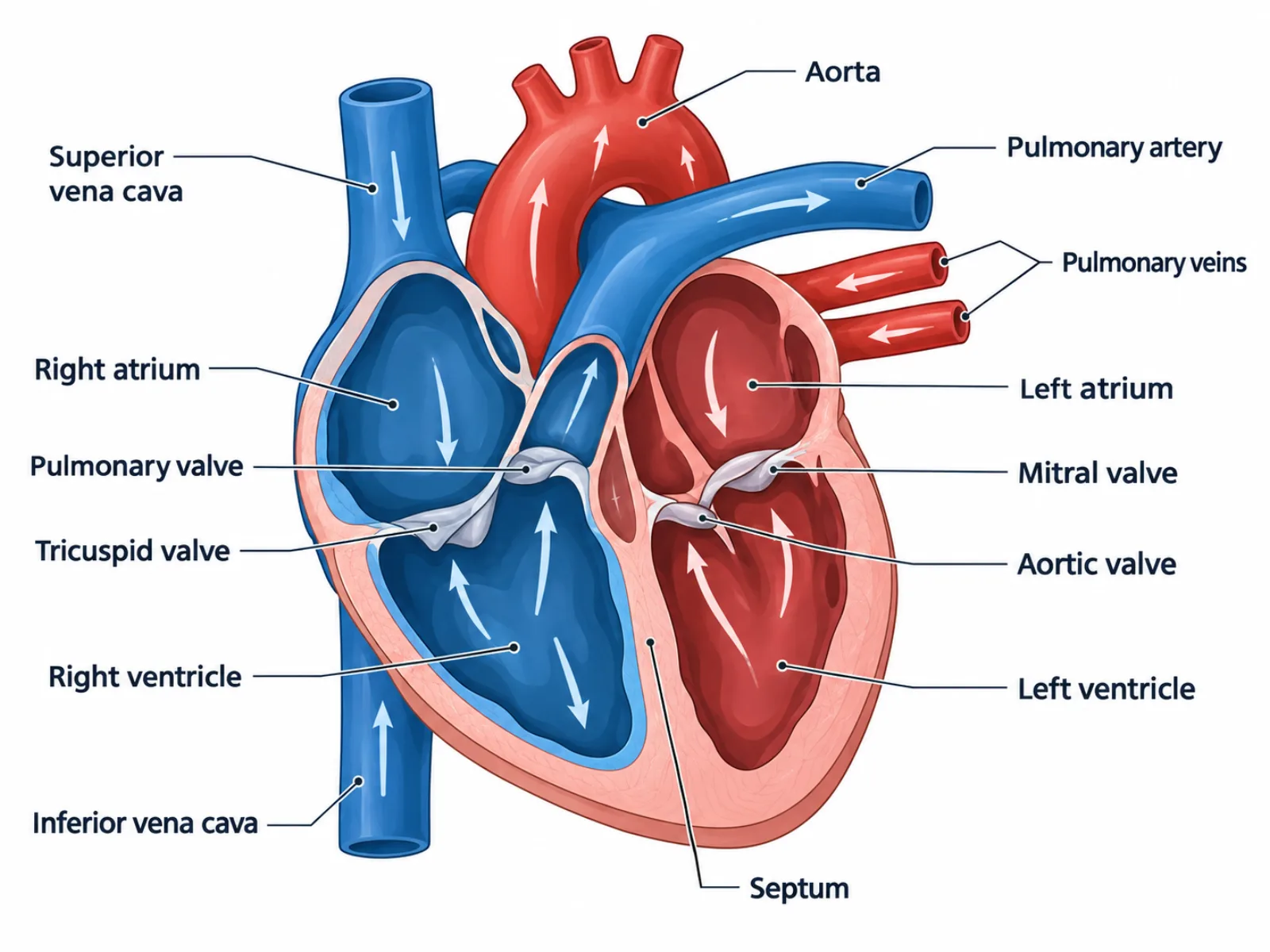
A heart diagram is a cross-sectional illustration that shows the four chambers, four valves, and major vessels of the human heart, along with the direction blood travels through them. Unlike a photograph, a labeled heart diagram cuts the organ open so you can see structures that would otherwise be hidden inside muscle, and it uses arrows to show the sequence of blood flow rather than just static anatomy. Because the heart is really two pumps working side by side — one for the lungs, one for the body — a good heart diagram has to represent both loops clearly without letting the viewer mix them up.
What a Heart Diagram Represents
At its core, a heart diagram is a map of two circuits sharing one muscular pump. The right side of the heart collects blood that has already delivered its oxygen to the body and sends it to the lungs for a refill; the left side receives that freshly oxygenated blood and sends it out to every organ and tissue. A diagram makes this division visible by drawing the septum as a clear vertical wall and by coloring or labeling the right-side structures separately from the left-side ones. Reading the diagram correctly means recognizing that blood never crosses from one side to the other except by passing through the lungs first.
The Labeled Parts of a Heart Diagram
- Right atrium — receives deoxygenated blood returning from the body via the superior and inferior vena cava.
- Tricuspid valve — a three-flap valve between the right atrium and right ventricle that prevents blood from flowing backward when the ventricle contracts.
- Right ventricle — pumps deoxygenated blood through the pulmonary valve into the pulmonary artery, sending it to the lungs.
- Pulmonary valve — prevents blood from flowing back into the right ventricle after it is ejected toward the lungs.
- Pulmonary artery — carries deoxygenated blood from the right ventricle to the lungs, the one artery in the body that does not carry oxygen-rich blood.
- Pulmonary veins — return oxygenated blood from the lungs to the left atrium, the counterpart exception where a vein carries oxygenated blood.
- Left atrium — receives oxygenated blood from the pulmonary veins.
- Mitral (bicuspid) valve — the two-flap valve between the left atrium and left ventricle.
- Left ventricle — the chamber with the thickest wall in the entire heart, pumping oxygenated blood through the aortic valve into the aorta and out to the whole body.
- Aortic valve — prevents backflow into the left ventricle once blood is pushed into the aorta.
- Aorta — the body's largest artery, distributing oxygenated blood to every organ system.
- Septum — the muscular wall separating the right and left sides, keeping oxygenated and deoxygenated blood from mixing.
- Superior and inferior vena cava — the two large veins delivering deoxygenated blood from the upper and lower body into the right atrium.
How to Read the Diagram: Following the Blood Flow
The most reliable way to interpret a heart diagram is to trace a single drop of blood through the entire circuit rather than trying to memorize all the labels at once. Start at the superior or inferior vena cava, follow it into the right atrium, through the tricuspid valve into the right ventricle, out through the pulmonary valve and pulmonary artery to the lungs — this is the pulmonary circulation loop. The same drop then returns through the pulmonary veins into the left atrium, through the mitral valve into the left ventricle, and out through the aortic valve and aorta to the body — the systemic circulation loop. Most diagrams draw arrows along this path, and the arrows matter more than the static shapes: they tell you which valve opens in which direction and confirm that flow is always one-way. Pay attention to wall thickness too — a diagram that renders the left ventricle wall visibly thicker than the right is being anatomically accurate, not just stylistic, since that thickness reflects the higher pressure needed to pump blood throughout the body.
Common Mistakes When Reading or Drawing a Heart Diagram
The single most common error is flipping left and right, because the heart is drawn from the perspective of the person it belongs to, not the viewer looking at the page — the chambers labeled "left" appear on the right side of the image. A second frequent mistake is assuming the pulmonary artery must carry oxygenated blood because it is an artery; students often apply the general rule "arteries carry oxygenated blood, veins carry deoxygenated blood" without realizing the pulmonary vessels are the deliberate exception, since they connect the heart to the lungs rather than to the rest of the body. A third mistake is misplacing the valves, particularly forgetting that each valve sits at the exit of an atrium or ventricle rather than floating freely inside a chamber — the tricuspid and mitral valves guard the atrium-to-ventricle openings, while the pulmonary and aortic valves guard the ventricle-to-artery openings.
Systemic vs. Pulmonary Circulation Within the Diagram
A single heart diagram actually encodes two separate circulatory loops, and distinguishing them is essential to reading it correctly. The pulmonary loop — right ventricle to lungs to left atrium — is short, low-pressure, and exists purely to oxygenate blood. The systemic loop — left ventricle to the aorta to the entire body and back to the right atrium — is long, high-pressure, and delivers oxygen and nutrients to every tissue. This is why the left ventricle wall is so much thicker than the right: it is built for the higher resistance of pushing blood through the entire body's vasculature, while the right ventricle only needs enough force to reach the adjacent lungs. Recognizing these as two connected but functionally distinct loops, rather than one continuous flow, is what separates a surface-level reading of a heart diagram from a functional understanding of how the heart actually works.